Fever without Localizing Signs and Symptoms
Authors: Christopher J Grace, MD, FACP, Brad Robinson, MPH, M.D.
Fever is a nonspecific physiologic response to inflammation. Infectious and non-infectious illnesses can present with fever mediated through the same cytokine pathways. Although most commonly attributed to localized infections, fever may be due to infections presenting non-focally (Table 1) or from a variety of non-infectious processes (Table 2).
Infectious Diseases Presenting Non-focally (Table 1)
A myriad of viral pathogens can cause febrile illnesses. Most present nonspecifically and may be both difficult to diagnosis and self limited in nature lasting days to a week or more. Many are associated with either upper respiratory infection or gastrointestinal symptoms. Patients, although uncomfortable, are non-toxic and do not require extensive diagnostic workups or empiric antibiotics. Other more serious infections can also present nonspecifically with a “flu-like” illness. Diagnosing these illnesses can initially be challenging and a high index of suspicion is needed especially those that do not have the more common upper respiratory infection or gastrointestinal symptoms. Several of the more serious and potentially life threatening infections are briefly reviewed below.
Infective Endocarditis
Infective endocarditis usually presents with an ill defined febrile illness. The clinical course can be rapid over days to weeks (Staphylococcus aureus) or more subacute over weeks to months (viridans streptococci). A heart murmur can usually be heard although the classic physical examination finding of a “new” murmur is uncommon. Peripheral stigmata of infective endocarditis such as petechiae, splinter hemorrhage, Osler nodes and Janeway lesions are also uncommon. Blood cultures (two sets) drawn before antibiotic therapy are very sensitive in isolating the pathogen and are key to the diagnosis. Infective endocarditis should be included in the differential diagnosis of a prolonged “influenza.”
Staphylococcus aureus Bacteremia
Staphylococcus aureus bacteremia is one of the most common and lethal community and hospital acquired bacteremias. Often associated with intravenous catheters or skin infections occurring in those with co-morbid medical illnesses, it can present non-focally in the otherwise healthy patient. Metastatic infection to heart valves, vertebral discs, joints and solid organs is common. Drainage of abscesses and prolonged intravenous antibiotic therapy is required.
Rocky Mountain Spotted Fever and Other Rickettsial Illnesses
Rocky mountain spotted fever, a tick borne illness caused by Rickettsia rickettsii, is endemic in the south eastern and south central United States. Patients present with fever, myalgia, headache, abdominal pain, nausea and vomiting. The characteristic maculopapular rash with its centripetal distribution begins between the second and fifth day of illness. With time the rash may become petechial or purpuric. 10-15% of patients do not develop a rash. Diagnosis is usually clinically based and confirmed by serology.
Coxiella burnetii (Q fever) a cause of atypical pneumonia may also present as a self limited febrile illness. Other rickettsial illnesses such as tick, louse borne or scrub typhus are usually associated with overcrowded hygienically poor environments or international travel.
Ehrlichiosis
Ehrlichiosis is a tick borne zoonosis. Human granulocytic anaplasmosis is caused by Anaplasma phagocytophilum while human monocytic ehrlichiosis is caused by Ehrlichia chaffeensis. Both are characterized by fever, headache, myalgias, thrombocytopenia, leukopenia and elevated serum transaminases. A maculopapular rash can occur but is relatively uncommon. Clinical illness can range from a mild flu-like illness to sepsis with renal and respiratory failure and central nervous system involvement. Diagnosis is most often made by polymerase chain reaction (PCR) amplification and serology.
Viral Infections
Hepatitis from hepatitis A, B or C virus may be asymptomatic or present with fever, myalgia, malaise and arthralgia. Jaundice and right upper quadrant discomfort may be present. Diagnosis is based on elevated transaminases and serologic testing.
Epstein-Barr Virus (EBV) is the causative agent of infectious mononucleosis. Infectious mononucleosis presents with fever, fatigue, pharyngitis and lymphadenopathy. Laboratory evaluation reveals elevated transaminases and atypical lymphocytosis. Diagnosis is based on detection of heterophile antibodies (mono spot test). Cytomegalovirus, Human Herpes Virus 6 (HHV-6) and Toxoplasma gondii can also cause a heterophile negative mono-like illness.
Primary infection with human immunodeficiency virus (HIV) can also cause a heterophile negative mono-like illness. Patients may present 2-6 weeks after exposure to the virus with fever, fatigue, weight loss, pharyngitis, myalgia,diarrhea, lymphadenopathy and headache. A maculopapular rash is present in 40-80% of patients. Early during acute seroconversion, the HIV antibody titer may be non-detectable. Diagnosis can be confirmed by repeat antibody testing and by detection of HIV RNA by plasma PCR (viral load).
Malaria and Travel Related Illnesses
A travel history should be obtained in all patients with unexplained fever. Malaria presents non-specifically with fever, headache and myalgia often accompanied with nausea, vomiting and diarrhea. The classic tertian or quartan fever patterns are rarely evident in the returning traveler. There may be anemia, leukocytosis or leukopenia and thrombocytopenia. Diagnosis is confirmed with the use of thick and thin blood smears reviewed by an experienced microbiologist. At least three smears should be performed before the diagnosis is considered ruled out. Other nonlocalizing febrile illnesses that should be considered in the returning international traveler include dengue, typhus,typhoid, leptospirosis and viral hepatitis.
Infections in the Elderly
The elderly, especially those living in nursing homes, are more prone to infections and those infections are associated with higher mortalities as compared to the young. Classic presentations of routine infections may not occur in the elderly and fever may not be present. Symptoms attributed to the infected organ may not be present; the elderly patient with pneumonia may not experience cough or complain of shortness of breath. Instead the elderly patient may present with change in functional status, worsening cognition, lethargy, falls and incontinence. The elderly patient with change in functional status should be evaluated for an infection even if they are afebrile and do not having localizing symptoms or signs of infection.
Non-infectious Causes of Fever (Table 2)
Drug Fever
Drug reactions can present with rash, fever, and/or eosinophilia, although all three symptoms occur in only a minority of cases. A previous history of drug allergy is present in less than 10% of the patients. The onset of a drug reaction occurs after an average of 7 days of drug use though drug reactions may occur as long as three weeks after initiation of the offending agent. No distinguishing fever pattern or temperature height helps to differentiate drug fever from other infectious or non-infectious causes of fever. Temperatures greater than 103o (F) can occur along with shaking chills in over half of the patients. Relative bradycardia is uncommon. The rash, if it occurs, is a pruritic maculopapular erythema covering most of the body. It has been reported in less than 20% of patients with a drug reaction. Leukocytosis can be seen. Eosinophilia is reported in 22% of patients but is generally mild and does not correlate with the severity of the reaction. Once the implicated drug is discontinued, the fever almost always resolves within 24-36 hours. Agents that can cause drug fever are listed in Table 3. Sympathomimetic agents such as epinephrine, cocaine and amphetamines can cause temperature elevation. Large doses of anticholinergic agents such as atropine, trihexyphenidyl or benztropine mesylate may also cause fever. Amphotericin B and bleomycin can act as pyrogens causing fever during or shortly after administration. The Jarisch-Herxheimer reaction is a febrile reaction caused by the bacteriocidal effect of penicillin on Treponema pallidum during the treatment of syphilis. Chemotherapy-induced tumor cell lysis can result in febrile reactions. Malignant hyperthermia and neuroleptic malignant syndrome are other drug induced causes of fever. See the Fever Chapter. Acute hemolysis and fever can occur in the glucose-6-phosphate dehydrogenase deficient individual exposed to sulfonamides, antimalarials, nitrofurantoin, quinidine and chloramphenicol.
Malignancy
Fever may be a presenting symptom or part of a symptom complex in a patient presenting with malignancy. The most common malignancies that can cause fever include lymphoma, acute leukemia, hepatocellular carcinoma, renal cell carcinoma and various solid tumors with metastasis to the liver.
Malignant lymphoma, including Hodgkin’s Disease and Non-Hodgkin’s Lymphoma, is the most common cancer to cause fever. It is present in as many as 25-30% of patients with Hodgkin's disease and less than 20% in Non-Hodgkin’s lymphoma. The classic, but infrequent, Pel-Ebstein fever of Hodgkin’s disease is a pattern of relapsing episodes of evening fevers that last for 3 to 10 days alternating in cyclic fashion with afebrile periods.
Fever is the presenting symptom in 10% of patients with acute myelogenous leukemia, although a portion of these will have associated neutropenic fever related infection that is actually causing the fever.
Hepatocellular carcinoma or hepatoma generally occurs in the setting of underlying cirrhosis especially if caused by chronic hepatitis B or C virus infection. Hepatic metastasis from solid tumors such as from breast, lung, and gastrointestinal cancers can occasionally cause fever.
Renal cell carcinoma most often presents with hematuria or flank pain, although fever occurs in about 20% of patients. The persistently febrile patient with hematuria in whom a urinary track infection is not clearly the source should be suspected of having renal cell carcinoma.
Collagen Vascular Disease
Still’s disease is a disorder of unknown etiology characterized by seronegative polyarthritis, fever and rash. The illness has a bimodal age distribution. The first peak, termed “systemic-onset juvenile rheumatoid arthritis”, occurs in childhood. The second peak, labeled “adult-onset Still’s disease”, occurs in the third or fourth decade. The rash is evanescent, salmon-colored, macular or maculopapular, nonpruritic, and typically occurs over the neck, trunk and extensor aspects of extremities. There is often a leukocytosis and marked elevation of the erythrocyte sedimentation rate (ESR).
Systemic lupus erythematosus is a multisystem disease characterized by a rash, arthritis, polyserositis, fever, oral or nasal ulcerations and fatigue. Most patients with fever caused by systemic lupus erythematosus show clinical evidence of active disease affecting multiple organ systems associated with leukopenia and high titers of antinuclear antibody.
Temporal arteritis, also known as cranial or giant cell arteritis, is a disease of the elderly characterized by headache, scalp tenderness, thickened tender or pulseless temporal artery, jaw claudication, fever, anemia, and markedly elevated ESR. However, it can often present subacutely with fever and nonspecific constitutional symptoms. The occurrence of temporal arteritis is closely associated with polymyalgia rheumatica. The ESR is usually > 50 in over 80% of cases.
The vasculitis is a heterogeneous group of illnesses characterized by inflammation of the blood vessels. Vasculitis can exist as a primary disorder or as a secondary manifestation of infections (hepatitis B and C virus, Epstein-Barr virus, infective endocarditis), rheumatologic diseases (systemic lupus erythematosus, rheumatoid arthritis, Sjögren’s syndrome), and certain malignancies (hairy cell leukemia). Table 4 summarizes the better defined primary vasculitic syndromes.
Miscellaneous
Acute Rheumatic Fever is a sequela of Streptococcus pyogenes pharyngitis. The antecedent pharyngitis is asymptomatic in about half of the patients. All patients demonstrate serologic evidence of recent S. pyogenes infection. The latent period between pharyngitis and onset of acute rheumatic fever averages 18 days. Patients may present with carditis, polyarthritis, chorea, subcutaneous nodules and/or erythema marginatum. Fever is one of the minor Jones Criteria.
Sarcoidosis is a chronic multisystemic disease of unknown etiology characterized by noncaseating granuloma formation primarily in the lungs and lymph nodes. It is more prevalent in women and African-Americans. It typically presents in persons 20-40 years of age with bilateral hilar lymphadenopathy, pulmonary infiltrates, and ocular and dermatological manifestations. Fever, if present, is usually seen in association with erythema nodosum, polyarthralgia and hilar lymphadenopathy.
Crohn’s Disease and ulcerative colitis are idiopathic inflammatory bowel diseases typically presenting with diarrhea and abdominal pain often associated with fever, fatigue, anorexia and weight loss. Some patients, particularly the elderly, may present with unexplained fever.
Pulmonary embolism most often presents as the sudden onset of pleuritic chest pain, shortness of breath and hemoptysis. Fever may be associated with these symptoms and rarely be the presenting symptom. Deep venous thrombosis can occasionally cause fever in addition to the more usual symptoms of pain, swelling and erythema often suggestive of cellulitis.
Hyperthyroidism can produce fever through excess thyroid hormone altering thermoregulation. Thyroiditis, an inflammatory disorder of the thyroid, can cause both cytokine release and thyroid hormone leakage from the injured gland to incite fever.
Gout is an acute inflammatory arthritis due to deposition of sodium urate crystals in the joints. The affected joint or joints are painful, swollen and erythematous and often are clinically indistinguishable from a septic arthritis. Fever may accompany the acute arthritis. In severe polyarticular attacks, high fevers and systemic toxicity are not unusual.
Approach to the Febrile Patient
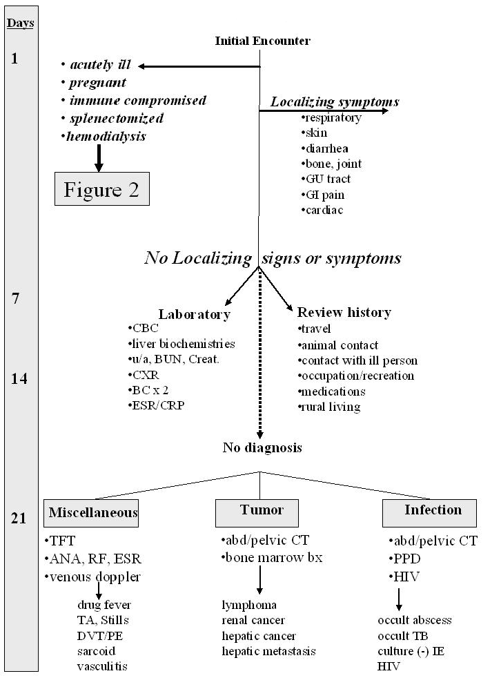
The approach to the febrile patient must be systematic and accomplished in a step-wise manner. See Figure 1. Many fevers are brief (3-7 days), viral in nature, and self-limited. The initial patient encounter should involve a history and physical examination to rule out life threatening illnesses and to triage patients who need a more thorough evaluation. The degree of illness of the patient and co-morbid conditions need to be taken into account when making decisions concerning rapidity of evaluation and need for empiric antibiotics.
For most patients, if the initial history and physical is unrevealing or suggests a viral respiratory tract infection, watchful waiting is appropriate. Any localizing symptoms or signs that suggest a discrete infection (e.g. urinary tract infection, cellulitis, arthritis or pneumonia) should be pursued with appropriate diagnostic tests and therapy.
If fever persists in the non toxic appearing patient for greater than one week without obvious source, then further evaluation with complete blood count (CBC), urinalysis, liver biochemistries, two sets of blood cultures, and a chest x-ray is warranted. If fever persists after 1-2 weeks and initial laboratory assessment is not revealing, a more exhaustive history (Table 5) and repeat physical examination should be performed. In general, therapeutic trials (especially of antibiotics) should be avoided since they only obscure the diagnosis. If the fever continues without resolution or diagnosis, it evolves into the more classic Fever of Unknown Origin category.
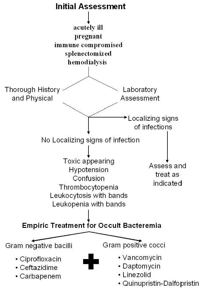
Patients who appear toxic with unstable vital signs, who are immunocompromised or pregnant, who have had a splenectomy or who are receiving hemodialysis should have a more rapid and thorough examination and immediate laboratory assessment including a CBC, liver biochemistries, blood urea nitrogen and serum creatinine, urinalysis, two sets of blood cultures and a chest x-ray. See Figure 2. Tachycardia, tachypnea, orthostatic blood pressure changes, altered mental status, purpuric skin lesions, thrombocytopenia, leukopenia or leukocytosis associated with increased band forms and increased serum creatinine raise the concern for occult bacteremia and sepsis. In these ill patients, empiric antibiotics should be initiated based on the microbiology from the presumed site of infection. If the ill appearing or septic patient has no localizing symptoms or signs, then the initial antibiotic empiricism should be directed towards an occult bacteremia. Antibiotics should coverStaphylococcus aureus (including methicillin resistant), streptococci and aerobic gram negative bacilli including Pseudomonas species. Initial regimens may include vancomycin, daptomycin, linezolid or quinupristin - dalfopristin for the gram positive bacteria and ciprofloxacin, ceftazidime or a carbapenem for the gram negative bacilli. Reassessment of the infectious illness and antibiotic selections can be made based on results of the blood cultures.
Fever of Unknown Origin
Fever of unknown origin has been defined as a temperature of greater than 38.3oC (101 F) for more than 3 weeks duration despite an intensive diagnostic evaluation over a week’s time. Classic fevers of unknown origin fall into infectious, malignant, vasculitic/rheumatologic and miscellaneous categories. See Table 6. Nosocomial fever of unknown origin is defined as fever starting more than 72 hours after hospital admission. See Table 7. Neutropenic fever of unknown origin has been defined as a temperature greater than 101o (F) along with an absolute neutrophil count of < 500 cells/mm3. See Table 8. HIV related fever of unknown origin is defined as four weeks of fever in the outpatient setting. These patients are usually very immunosuppressed with CD4 lymphocyte counts < 100 cells/mm3. See Table 9. Newly infected patients may present with mononucleosis like illness with fever, hepatitis, aseptic meningitis and rash.
Fever of unknown origin is much more often due to an atypical presentation of a common disease than to a typical presentation of an uncommon one, i.e., a patient with a diverticular abscess without abdominal pain vs. Still’s disease. For the patient with a prolonged cryptic fever, the importance of HIV testing cannot be overemphasized.
It is the epidemiological setting in which the patient presents (Table 10 and 11) and clues from the history (Table 5) and laboratory investigation (Table 12) that suggests the focus of investigation.
Empiric therapy is discouraged since it is rarely curative and has the potential to delay diagnosis. Supportive therapy with nonsteroidal anti-inflammatory drugs (NSAIDs) while observing for the evolution of clues appears to be a safe approach. Only very rarely do patients deteriorate on NSAIDs without presenting new diagnostic clues. Consultation with infectious disease specialists is encouraged.
Tables
Figure 1: Approach to the Febrile Patient
The evaluation of the febrile patient is paced according to severity of symptoms, underlying illnesses and results of history, physical exam and laboratory assessment. It should proceed in a stepwise fashion from non-invasive inexpensive testing to the more complex evaluation frequently needed in the evaluation of the patient with FUO. The acutely ill or immune compromised patient, pregnant women, and the patient receiving dialysis or living in a nursing home should receive a more in-depth initial investigation. The shaded box on the left side represents the number of febrile days.
ICH= immune compromised host, IE= infective endocarditis, CXR=chest x-ray, CAT=computer assisted tomography, TB=tuberculosis, TFT=thyroid function tests, DVT=deep venous thrombosis, PE= pulmonary embolism, TA=temporal arteritis, GI=gastrointestinal, GU=genitourinary, BC=blood culture, u/a=urinalysis, CA=cancer, CBC=complete blood count. Adapted form Marcel Dekker Inc in Medical Management of Infectious Diseases, Ed C Grace, 2003.

Figure 2: Empiric Antibiotics for Ill Appearing Patient at Risk for Occult Bacteremia

Table 1: Infections That May Present Non-focally
| Infectious endocarditis |
| Staphylococcus aureus bacteremia |
| Rocky mountain spotted fever and other rickettsial illnesses |
| Ehrlichiosis |
Viral infections:
|
| Malaria and travel related infections |
| Infections in the elderly |
Table 2: Non-infectious Illnesses That May Present with Fever
| Drug fever |
Malignancies:
|
Collagen vascular diseases:
|
Miscellaneous:
|
Table 3: Drugs That Can Cause Fever
| Antimicrobial | Cardiovascular | Anti-inflammatory | Central nervous system | Anti-neoplastic | Miscellaneous |
|---|---|---|---|---|---|
•beta lactams: ·penicillins ·cephalosporins •sulfonamides •TMP-SMX •amphotericin b •tetracycline •macrolides •streptomycin •vancomycin •isoniazid •para-aminosalicylic acid •nitrofurantoin •mebendazole |
•quinidine •procainamide •hydralazine •methyldopa •nifedipine •triamterene |
•salicylates •ibuprofen •tolmetin |
•carbamazepine •phenytoin •barbiturates •chlorpromazine •haloperidol •thioridazine •amphetamine |
•bleomycin •asparaginase •daunorubicin •procarbazine •cytarabine •streptozocin •6-mercaptopurine •chlorambucil •hydroxyurea |
•allopurinol •antihistamine •iodide •cimetidine •levamisole •metoclopramide •clofibrate •folate •prostaglandin e2 •ritodrine •interferon •streptokinase |
Used with permission by Marcel Dekker Inc in Medical Management of Infectious Diseases, Ed C Grace, 2003.
Table 4: Vasculitis Syndromes
Syndrome |
Population at risk |
Symptoms |
Organs involved |
Laboratory assessment |
|---|---|---|---|---|
Polyarteritis nodosa |
middle-aged men>women |
fever, wt loss, rash, abdominal pain, arthralgia |
kidneys, GI tract, skin, peripheral nerves |
leukocytosis anemia, ↑ESR, p-ANCA, HbsAg, U/A |
Churg-Strauss
|
middle-aged men>women |
fever, wt loss, rash, asthma |
lungs, skin, peripheral nerves |
eosinophilia, p-ANCA, abnormal CXR |
Wegener’s granulomatosis |
male or female, adolescent to middle aged |
fever, wt loss, nasal ulcers, cough, sinusitis, hemoptysis, arthralgia |
upper and lower respiratory tracts, kidney |
Leukocytosis, anemia, ↑ESR c-ANCA, U/A, abnormal CXR |
Takayasu’s arteritis |
young female, more common in Orient |
fever, wt loss, arthralgia, loss of peripheral pulses, pain over vessels |
aortic arch and branches |
arteriography |
Henoch Schönlein purpura |
children and young adults |
palpable purpura, arthralgia, abdominal pain, bloody stool |
skin, kidneys, GI tract |
U/A |
Essential mixed cryoglobulinemia |
middle aged women |
purpuric lesions, ulcers, arthralgia, Raynaud’s phenomenon |
kidney, skin |
low C3, C4, CH50, U/A, HCV antibody |
ESR = erythrocyte sedimentation rate, ANCA = antinuclear cytoplasmic antibody, U/A = urinalysis, HbsAg = hepatitis B virus surface antigen, CXR = chest x-ray, GI = gastrointestinal, C = complement, HCV = hepatitis C virus
Table 5: Key Historical Questions
Medical history:
• malignancies • organ transplantation
|
| Recent contacts with persons having similar illness |
| Recent and past travel, places of residence and military service |
| Animal exposures at home, work or recreational |
| Work exposures |
| Recreational exposures including rustic living arrangements, animals, tick bites |
| Unusual dietary habits |
| TB exposure |
History of high risk behavior:
|
| History of transfusions, immunizations |
| Complete list of medications including over the counter and “alternative” remedies. |
| Drug or other allergies |
| Ethnic origin and familial history of fevers, tuberculosis, collagen-vascular diseases, cancer, thrombosis, anemia |
| Living in a rural area |
Table 6: Causes of “Classic” FUO by Etiologic Category
| Infection | ||
|---|---|---|
| Localized bacterial: | Systemic bacterial : | Viral: |
|
|
|
| Systemic mycoses: | Endovascular: | Parasitic: |
|
|
|
| Neoplasm | ||
| Fever relatively common: | Occasional cause of fever: | |
|
|
|
| Non-Infectious Inflammatory Conditions | ||
| Rheumatologic: | Vasculitis: (Table 4) | Granulomatous diseases: |
|
|
|
| Thromboembolism: | Endocrine: | Fever: |
|
|
|
CMV = cytomegalovirus, EBV = Epstein Barr Virus, HAV = hepatitis A virus, HBV = hepatitis B virus, HCV = hepatitis C virus, HIV = human immunodeficiency virus, SLE = systemic lupus erythematosus, DVT = deep venous thrombosis, PE = pulmonary embolism
Table 7: Causes of “Nosocomial” FUO by Etiologic Category
Nosocomial infections:
|
| Drug fever (Table 3) |
| Thromboembolic disease |
| Alcohol, barbiturate, benzodiazepine, narcotic withdrawal |
| Blood product transfusion |
| Pancreatitis |
| Phlebitis |
| Gout |
| Acute myocardial infarction and Dressler’s syndrome |
| Post-operative inflammation |
Table 8: Causes of “Neutropenic” FUO by Etiologic Category
Infections:
|
Drug fever
|
| Malignancy related fever |
| Thromboembolic disease |
| Blood product transfusion |
| Intravenous catheter related phlebitis |
Table 9: Causes of “HIV” FUO by Etiologic Category
|
Table 10: Differential Diagnosis of Cryptic Fever by Age
| Fever in the younger adult: | Fever in the elderly adult: |
|---|---|
|
|
EBV = Epstein Barr Virus, CMV = cytomegalovirus, HIV = human immunodeficiency virus, SLE = systemic lupus erythematosus, UTI = urinary tract infection
Table 11: Infectious Etiologies of Fever in the Patient with Animal Contact
Infection |
Pathogen |
Animal exposure |
Transmission |
Clinical signs and symptoms |
Diagnosis |
|---|---|---|---|---|---|
Brucellosis |
Brucella Melitensis B. abortus B. suis |
goats, sheep cattle hogs |
unpasteurized milk or cheese, contaminated meat,aerosolized animal fluids |
fever, chills, arthralgias, lymphadenopathy HSM, epididymoorchitis |
Serum antibodies |
Tularemia |
Francisella tularensis |
wild rabbits, small rodents |
direct contact with infected tissues, inhalation of aerosol, tick bite |
ulcero-glandular ocular-glandular pneumonia, typhoidal fever and prostration |
Serum antibodies |
Leptospirosis |
Leptospira interrogans |
rats dogs cattle, pigs |
drinking or swimming in contaminated water |
flu-like illness then meningitis, hepatitis, hematuria |
Isolation of spirochete from urine, blood, CSF, serum antibodies |
Q fever |
Coxiella burnetii |
cattle, sheep, goats |
infected aerosol from parturient animals, ingesting contaminated milk |
flu-like illness, pneumonia, hepatitis. no rash. |
Serum antibodies |
Psittacosis |
Chlamydia psittaci |
birds |
infected aerosol from bird excreta |
flu-like, splenomegaly, pneumonia |
Serum antibodies |
CSF = cerebrospinal fluid
Table 12: Laboratory Clues to the Etiology of Fever
| Lymphocytosis: | Atypical lymphocytosis: | Eosinophilia: |
|---|---|---|
|
|
|
| Monocytosis: | Leukopenia: | Lymphopenia: |
|
|
|
| Thrombocytopenia: | Elevated ESR: | Alkaline phosphatase: |
|
|
|
| Elevated transaminases: | Abnormal urinalysis: | |
|
|
|
GUIDED MEDLINE SEARCH FOR:
Reviews
Gilbert DN. Use of Plasma Procalcitonin Levels as an Adjunct to Clinical Microbiology. J Clin Microbiol 2010;48:2325-2329.
Bryan CS, et al. Fever of Unknown Origin: Is There a Role for Empiric Therapy? Infect Dis Clin N Am 2007;21:1213-1220.

