
Fever in the Postoperative Patient
Authors: Nikkiya M. Fraser, M.D., Julie E. Mangino, M.D.
INTRODUCTION
Postoperative fever presents a frequent and at times, thorny issue for the clinician. While fever is usually a normal phenomenon in the immediate post-surgical period, vast amounts of resources are expended every day in the pursuit of more ominous diagnoses (1).
Normal body temperature encompasses a wide range of values, but for practical purposes a fever has been defined as 38.3oC (100.4o F) and above in ICU patients (1) and can be defined as such in surgical patients. In the first 48 hours of the postoperative period a fever is nearly always non-infectious in origin (2). The inflammatory mechanisms responsible for postoperative fever have been the subject of a number of studies. Tissue damage alone results in the disruption of phospholipids from the cell membrane, leading to a cascade of prostaglandins and cytokines which eventually lead to a body temperature elevation (3). However, fever that persists beyond 96 hours generally warrants further attention.
Similar to fever in the critically ill patient, a systematic approach to fever in the postoperative patient includes considering both the most common diagnoses in this group as well as ruling out those conditions associated with morbidity and mortality. Postoperative infections include device-related complications such as hospital or ventilator-associated pneumonia, catheter-related urinary tract infection, catheter-related bloodstream infection, and surgical site infections. Other, rarer, infections can include sinusitis, odontogenic infections, occult abdominal abscesses, or infectious sources that were incubating prior to the surgery but not identified by the surgical team. Non-infectious etiologies, such as deep venous thrombosis (DVT) must also be considered.
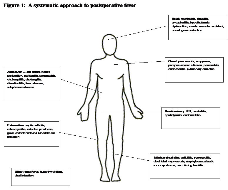
When developing a differential diagnosis for postoperative fever, a head-to-toe approach can be useful (Figure 1). Certain etiologies become more important depending on the extent of the surgical procedure and the postoperative day that the fever develops (Table 1).

INFECTION IN THE POSTOPERATIVE PATIENT
Nosocomial Pneumonia
Hospital-associated pneumonia (HAP) is the second most common nosocomial infection after urinary tract infection and is a frequent cause of infection in the postoperative patient (4). Surgical patients are at particular risk for the development of nosocomial pneumonia as many surgical procedures require general anesthesia and intubation. Intubation increases the risk of developing ventilator-associated pneumonia (VAP) by promoting aspiration and colonization of the endotracheal tube with endogenous and multi-drug resistant, healthcare-associated pathogens especially as the duration of intubation increases. Prompt weaning from mechanical ventilation decreases the risk of VAP (4). While many surgical patients require a relatively short period of ventilation compared to critical care patients, they are also at risk of developing pneumonia because of immobility, which promotes atalectasis and poor clearance of secretions.
If hospital-associated pneumonia is suspected, a chest x-ray is recommended to determine the severity of potential pulmonary involvement and to assess if more complicated processes, such as pleural effusion are present. The absence of an infiltrate on chest x-ray does not preclude the diagnosis of HAP. All patients with suspected HAP should also, ideally, have sampling of the lower respiratory tract by an endotracheal aspirated specimen, mini-broncho-alveolar lavage (BAL), or bronchoscopic BAL. Quantitative sputum cultures are generally more accurate than semi-quantitative. An assessment to rule out Legionella including urinary antigen detection would also be prudent. Recommendations for the management of HAP are outlined in Table 2.
Catheter-Related Urinary Tract Infection
Urinary tract infection (UTI) is the most common nosocomial infection overall, making up about 40% of all hospital-acquired infections (5). Nearly all patients will have a urinary catheter placed during and shortly after surgery, making this a common source of contamination and infection. Catheterization in itself is a risk factor for developing bacteriuria, and the risk of bacteriuria increases with the duration of catheterization. Most cases of bacteriuria are asymptomatic; however, up to 30% of cases may be complicated by fever or other symptoms (6). Fever is more common in long-term catheterization than in the short term. Fever in a catheterized patient could also be indicative of complications such as:pyelonephritis, prostatitis, epididymitis, or urethritis (7).
Since asymptomatic bacteriuria is common in catheterized patients, catheterized urine cultures are not necessary or useful unless there are symptoms present, which may or may not be assessable based on the patient’s level of sedation. A positive urine culture accompanied by pyuria (demonstrated by urinalysis) is much more helpful in the febrile patient. Pyuria is defined as ≥10 white blood cells/µL or ≥3 white blood cells/high-power field of unspun urine. A urine culture with ≥105 cfu/mL with no more than two species of organisms is also considered to be more suggestive of a true UTI (5).
Candiduria is a frequent finding among hospitalized patients and most are asymptomatic. Yeast in the urine most commonly represents either colonization of the urinary catheter or perineum and rarely leads to candidemia unless obstruction is present or instrumentation of the urinary tract is performed. No well-defined criteria exist for defining Candida UTI in terms of pyuria or quantitative urine cultures. The Infectious Diseases Society of America has recommended that patients with neutropenia, renal transplants, symptomatic patients, or those undergoing genitourinary procedures be treated if they develop candiduria. Candiduria in a patient with neutropenia can be a marker for disseminated infection. However, overwhelming evidence suggests that candiduria as an isolated observation does not portend to subsequent invasive disease. Therefore, it is less imperative to use antifungal agents to treat patients who have candiduria (8).
Surgical Site Infection
Surgical site infections (SSIs) have been estimated to be the most common nosocomial infection among surgical patients (9), occurring in approximately 3% of all surgical procedures (10). A SSI is currently defined as an infection arising within 30 days of an operative procedure and at the site of surgical intervention, if no implants were placed (5). Most SSIs develop within 5 to 10 days after a procedure, although some may develop up to 30 days or later (10).
In the majority of SSIs, pathogens are introduced at the time of the operative procedure. These pathogens usually originate from the patients’ own skin or bowel flora.
The risk of developing a SSI depends on the patient’s overall state of health and the type of procedure performed. Surgical procedures have been traditionally classified according to the anticipated risk of contamination (Table 3). The risk of developing an SSI has been shown to increase with the level of contamination in the wound. The National Nosocomial Infections Surveillance System (NNIS) has also developed a risk index (Table 4). Characteristics of the patient and the procedure which can effect the development of SSI are outlined in Table 5.
Surgical site infection should be suspected if there are signs of local inflammation, such as erythema, warmth, or tenderness. After further work-up or surgical re-exploration, the infection may be classified as either superficial: involving the skin and subcutaneous tissue alone, deep: involving the fascia and muscle, or an organ/space infection: involving organs deep to the incision, but directly related to the surgery.
Obtaining a culture from a SSI is especially problematic and prone to contamination with local flora. Wound cultures are not recommended for a suspected SSI unless signs of systemic toxicity are present such as fever or leukocytosis. If a culture is taken, an aspirate of pus in a syringe or a tissue specimen will provide the most meaningful information versus routine swabs of wound drainage (10).
Necrotizing soft-tissue infections are a rare but very serious subset of surgical site infections. Clostridial myonecrosis or gas gangrene is most frequently caused by Clostridium perfringens although other species of Clostridium have also been implicated. Clostridial myonecrosis can occur within a few hours of surgery and is characterized by severe pain that extends beyond the original wound. The surrounding tissue and muscle becomes necrotic resulting in skin discoloration and the formation of hemorrhagic bullae. Purulent drainage is often absent and a thin, serous discharge is more common. Necrotizing fasciitis caused by group A Streptococcus has also been reported in the postoperative period and may be associated with hypotension, renal failure, and respiratory failure. Both cases are considered surgical emergencies and rapid surgical intervention is needed to remove necrotic tissue. Antibiotics are secondary in importance to surgical resection, although parenteral penicillins and clindamycin are considered the treatment of choice and may have a role in halting the spread of infection (11).
Catheter-Related Bloodstream Infection
While most surgical procedures will only require the use of simple peripheral IV access, more involved procedures will require peripheral and nontunneled central venous catheters (CVCs), pulmonary artery catheters, and other intravascular devices. All of these devices provide a portal of entry for infection. About 200,000 nosocomial bloodstream infections occur in the United States yearly; nontunneled CVCs are implicated in the majority of these (12).
Catheter-related bloodstream infection should be considered in any febrile patient with a CVC; inflammation or purulence at the catheter site may not necessarily be present but strongly suggests this diagnosis when it is. If a catheter-related bloodstream infection is suspected, two sets of blood cultures (with at least one drawn percutaneously) should be obtained. If an alternate site of infection is not identified or if the patient is septic, the catheter should be removed and the tip can be sent for quantitative or semi-quantitative cultures. A diagnosis is confirmed if the patient has clinical manifestations of infection (fever, chills, and hypotension), at least one positive blood culture which does not contain typical skin flora when obtained percutaneously, and no other potential source of infection. Additional confirmation can include a positive semi-quantitative (≥15 cfu per catheter segment) or quantitative (≥102 cfu per catheter segment) catheter culture of the same organism obtained peripherally. Comparing blood cultures from a peripheral vein and a catheter for “time to positivity” has been found to correlate with quantitative cultures and has been useful in many institutions (12).
Many catheter-related infections are caused by coagulase-negative staphylococci, including S. epidermidis. Other typical pathogens include Staphylococcus aureus, enterococci (including vancomycin-resistant isolates), gram-negative bacilli such as Pseudomonas, and Candida spp. (13) Some species, such as Corynebacterium and Bacillus, are usually considered contaminants but may be significant if present in a series of blood cultures (1).
Other Infectious Causes of Fever
A rare surgical complication which may present in the first 48 hours following surgery is staphylococcal toxic shock syndrome (TSS). This condition had been associated with the use of highly absorbent tampons; however, there have been reports in the literature of TSS secondary to surgery. TSS is characterized by fever, hypotension, a diffuse macular rash, desquamation, confusion and oliguria. In TSS the wound typically appears remarkably benign in contrast to the patient’s symptoms. Although this syndrome has been reported after a wide variety of surgical procedures, it has been reported most frequently following nasal surgery and is more likely to be associated with nasal packing (14).
Intra-abdominal infection is another serious complication and tends to be caused by highly-resistant organisms when occurring after surgery. Fever following abdominal surgery, particularly when accompanied by abdominal pain suggests this diagnosis. CT scan is the most frequently used modality for identifying abdominal abscesses and, in certain cases, CT guidance can also be used to obtain abscess material for culture. Therapy can be guided by culture of any intra-abdominal fluid collection, but there are a number of empiric agents that would be most useful once the diagnostic material is obtained. At least 0.5 cc of fluid will have the best yield, and this material should be sent for both aerobic and anaerobic culture. Swabs are not appropriate as they do not provide sufficient material for anaerobic cultures (15). Acute acalculous cholecystitis is a rare complication following non-biliary surgery and has been reported following trauma, burns, and cardiac surgery. There are no preoperative markers associated with an increased risk of acalculous cholecystitis although previous series have shown that this complication often accompanies multiorgan dysfunction, prolonged mechanical ventilation, and large transfusion requirements. In one retrospective review, acalculous cholecystitis was reported in 7 of 996 patients that had undergone aortic reconstruction. These patients also exhibited other organ involvement such as renal dysfunction and respiratory failure (16). This diagnosis should be suspected in a critically ill patient with fever, hyperbilirubinemia, and elevated liver transaminases. Abdominal ultrasound or biliary scintigraphy are the preferred imaging modalities.
Sinusitis is in the differential diagnosis for unexplained fever in the ICU. Nosocomial sinusitis is often associated with nasotracheal or nasogastric intubation which compromises the normal drainage of the sinuses and promotes colonization with pathogens. Some patients may also have pre-existing sinusitis before surgery. Air-fluid levels in the sinuses can be detected by plain radiograph, but adequate studies are difficult to obtain with portable equipment. CT scanning has a higher diagnostic yield in critically ill patients. Sinus aspiration under sterile conditions should be performed for gram stain and culture if radiographic studies suggest sinusitis. Pseudomonas aeruginosa is responsible for the majority of cases of nosocomial sinusitis, although gram-positive bacteria and fungi can also be present (16).
Clostridium difficile colitis should always be considered when fever develops following antibiotic exposure. Diarrhea accompanied by leukocytosis is supportive of this finding and diagnosis is made by sending the stool for the toxin assay (22).
Non-Infectious Causes of Fever
Atelectasis is often implicated in postoperative fever, especially in the first 48 hours after surgery. The concept of atelectasis as a cause of fever in and of itself, however, has been called into question (17). Atelectasis may be more important in the postoperative period as a potential risk factor for the development of pneumonia. Serial chest x-rays will help differentiate atelectasis from pneumonia.
Withdrawal from alcohol or benzodiazepines can be a possible cause of fever. It is particularly useful to inquire about substance abuse when evaluating a postoperative fever, but especially important to be aware of preoperatively (18).
The perioperative period is typically a period of exposure to new medications such as bowel evacuative agents (neomycin, erythromycin, or metronidazole), preoperative antibiotic prophylaxis, sedatives, and anesthesia itself. Drug fever should always be considered in the workup of a postoperative fever. While every medication can potentially cause drug fever, several medications are more frequent offenders (Table 6). The presentation of drug fever can be quite varied. Characteristics traditionally associated with drug fever, such as relative bradycardia, are not usually present. In a systematic review of patients with drug fever, only 18% had a reported rash, only 22% had eosinophilia and relative bradycardia was reported in only 11 % (19). In this series there was also no clear association between atopic disease and the development of drug fever. Timing of drug fever is variable and can occur shortly after the initiation of therapy or after long periods on the same medication (20). Drug fever is largely a diagnosis of exclusion and infectious causes for fever must be ruled out. The diagnosis can be confirmed by discontinuing the offending agent; the fever generally subsides within 48 to 72 hours.
Malignant hyperthermia occurs in genetically susceptible patients exposed to certain anesthetics, mainly succinylcholine or halothanes. Most of these cases occur in the operating room but can also occur up to 24 hours afterward (18).
Endocrine abnormalities may be another source of fever. Surgery can precipitate thyroid storm in susceptible hyperthyroid patients. Acute adrenal insufficiency can be associated with fever and hypotension and have a presentation similar to septic shock. Acute adrenal insufficiency may occur in patients who take corticosteroids on a chronic basis and then have these medications rapidly withdrawn. This condition also develops during acute adrenal hemorrhage, secondary to disseminated intravascular coagulation or overuse of heparin or warfarin (1).
Deep venous thrombosis or pulmonary embolus should be considered as a cause of postoperative fever, particularly in the sedentary patient. Evaluation could include sonography of the lower extremities or sites of intravascular catheters and spiral CT of the chest.
APPROACH TO THE FEBRILE POSTOPERATIVE PATIENT
Key Points of History
Important points to review in the history and physical are outlined in Table 7. When presented with a patient with postoperative fever, one of the most important elements of the history is the surgical procedure itself. The operative report should always be reviewed and, if possible, the surgeon involved can be queried for additional details. Attention should be paid to the duration of the procedure and which, if any, perioperative antibiotics were used. One should note any use of “foreign material” such as mesh or grafts. Complications that occurred during the procedure, such as perforation of a viscus, should be addressed. The operative report will usually detail any material that was sent for culture or histological examination. All associated intraoperative cultures and surgical pathology reports should be reviewed.
The patient’s past medical history may also provide clues to determine the cause of a postoperative fever. The prior medical conditions are important, particularly the presence of diabetes mellitus. Prior surgeries should be reviewed, paying particular attention to whether they were complicated by postoperative infection or a long rehabilitation course. An evaluation for the presence of prosthetic joints, heart valves, pacemakers, ports, and other indwelling synthetic devices should be sought.
To rule out drug fever a medication history is particularly valuable. The home medications should be reviewed and compared with the in-hospital medication list, noting additions or deletions. The anesthesia record should be reviewed for perioperative antibiotics and other new medications given perioperatively such as: analgesics, DVT and stress ulcer prophylaxis therapy.
Many factors in the patient’s social history affect the postoperative course. Tobacco use has negative effects on wound healing and makes pulmonary complications more likely. Substance abuse, particularly intravenous substances, raises the possibility of other infectious complications, such as endocarditis or the diagnosis of HIV.
Key Points of Evaluation
The most logical place to start when evaluating the febrile postoperative patient is with the fever itself. While the fever curve is no longer used in fever diagnosis, it can still provide clues as to the cause of the fever. One example is with drug fever where fever spikes coincide with the administration of a scheduled medication. One should examine the patient’s temperature in relation to blood pressure, heart rate, and respiratory rate. Fever associated with tachycardia and hypotension may indicate a more serious underlying infection or the presence of bacteremia. Relative bradycardia or pulse-temperature dissociation is traditionally associated with drug fever.
Evaluation should include a careful physical exam, paying attention to the possible sources of infection. Observing the presence of devices (such as ventriculostomies) in the head and neck region and a gross evaluation for facial or sinus tenderness is a good starting point. The chest should be examined for rales or signs of consolidation. One should note respiratory effort and the presence of “splinting” which may hinder adequate ventilation and predispose to atalectasis and pneumonia. An incentive spirometer should be at the patient’s bedside and the patient can be asked to demonstrate its use. All intravascular devices, including peripheral IVs, should be examined for inflammation. The extremities should be examined for evidence of ischemia or DVT, such as cyanosis, edema, or tenderness. The skin should be examined for a rash, consistent with hypersensitivity reactions. A bed-bound patient should always be rolled so the back can be examined for skin breakdown, bullae or decubitus ulcers.
It is imperative to examine the wound itself after postoperative day 2, when it should be adequately granulated. All dressings and packing should be removed and the wound should be inspected for drainage, erythema, or purulence. The borders of the incision should be examined for signs of cellulitis and the presence of drains with the amount and character of the drainage noted.
Laboratory and radiological testing should be based on the physical findings and the most likely source of infection. Blood cultures should be collected in patients with fever and an intravascular device. Chest radiography can be performed on patients with suspected pneumonia. Fever and abdominal tenderness should prompt a CT scan of the abdomen. In the early postoperative period, however, post surgical inflammation may appear radiographically similar to early abscess formation. An ultrasound evaluation for DVT should also be considered.
Non-Antibiotic Management of Postoperative Fever
In the early stages of a postoperative fever, pulmonary toilet and incentive spirometry should be encouraged. Adequate pain control (without over-sedation) and removal of the urinary catheter can encourage ambulation to and from the bathroom and avoid tethering the patient to the bed.
NSAIDs in low doses can be useful in controlling fever in the immediate postoperative period. In one series, 15 mg of ketorolac given every 6 hours beginning in the recovery room following uterine fibroid removal was associated with a significant reduction in postoperative fever (3).
If bacteriuria is present, the urinary catheter should be removed. Most cases of catheter-associated bacteriuria clear with removal of the catheter and without additional antibiotics.
Indications for Antibiotic Therapy
Empiric Antibiotics
The most common pathogens implicated in postoperative infections are outlined in Table 8.
Nosocomial Pneumonia
With healthcare-associated pneumonia, antibacterial therapy should be started as soon as possible to reduce morbidity and mortality. The choice of empiric antibiotics depends on the patient’s pre-existing risk of infection with or without multidrug-resistant organisms (Table 2) as well as the local formulary and ICU antibiogram. Clinical improvement takes between 48-72 hours and the patient’s response should be re-evaluated after 3 days. Antibiotic coverage should be narrowed based on previously collected culture data or discontinued altogether if cultures are negative and fever has resolved. Specific antibiotics can be continued until treatment day 7 or 8, with an aim to finish antibiotics by day 14, according to current recommendations.
Catheter-Related Urinary Tract Infection
Most catheter-related UTIs are caused by invasion of the patient’s normal colonic flora. The most common pathogens in these cases are E. coli, Klebsiella pneumoniae, Proteus mirabilis, Enterobacter sp., enterococci, Staphylococcus aureus, and Staphylococcus epidermidis. Asymptomatic bacteriuria in the post-surgical patient does not require treatment, with the exception of patients who have undergone urologic surgery. For an uncomplicated UTI with associated fever, seven to ten days of antibiotic therapy is recommended (6).
Surgical Site Infection
Since most surgical site infections are caused by S. aureus, first-generation cephalosporins are commonly used for prophylaxis. Many antibiotics given perioperatively have relatively short half-lives and little to no post-antibiotic effect. A preoperative prophylactic antibiotic regimen may not have covered the causative organism for the SSI. Initial treatment of a superficial SSI is wound care before antibiotics and most inflammation at the wound site should resolve without any specific treatment. An infected wound is best managed by opening, draining any purulent material, and allowing it to heal by secondary intention. Antibiotics are indicated if there is evidence of systemic toxicity (fever, leukocytosis) or cellulitis extending more than 2 cm beyond the incision (10). The choice of empiric antibiotics depends on the type of surgery performed. For procedures that do not involve the gastrointestinal or female genitourinary tract,cefazolin, oxacillin, or coverage for MRSA is recommended (depending on the incidence of MRSA at a particular facility.) For gastrointestinal procedures, ampicillin/sulbactam, ertapenem, or a fluoroquinolones plus clindamycin are recommended (21).
For catheter-related bloodstream infections, the initial choice of empiric antibiotics will depend on local antibiograms. Many are caused by coagulase-negative staphylococci and Staphylococcus aureus and initial therapy should be aimed at these organisms. Vancomycin is usually recommended when there is an increased incidence of methicillin-resistant S. aureus and until susceptibilities are known (12). In immunocompromised or severely ill patients, a third or fourth-generation cephalosporin should be included to cover enteric gram-negative bacilli including Pseudomonas in addition to staphylococcal coverage.
Catheter-Related Bloodstream Infection
The duration of therapy for catheter-related bloodstream infections depends on the responsible organism and whether the infection is complicated or not. Bloodstream infections with coagulase-negative staphylococci and without metastatic sites of infection can be treated by removal of the catheter and 5-7 days of therapy. S. aureus infections can be treated for 14 days when the focus is removed and there are no metastatic sites of infection and TEE is negative. Most gram-negative bacilli and Candida spp. should be treated for 14 days (12). If septic thrombosis, endocarditis, or osteomyelitis develops because of the bloodstream infection, systemic antibiotics should be continued for at least 4-6 weeks (or 6-8 weeks in the case of osteomyelitis).
Other Infectious Causes of Fever
For suspected intra-abdominal infections, antibiotic therapy should be initiated as soon as possible. Empiric therapy should be guided by local resistance patterns. A potential regimen could include a b-lactam/b-lactamase inhibitor, a carbepenem, or a combination of a third or fourth-generation cephalosporin plus metronidazole (15).
Complications of Perioperative Antibiotics and Their Management
Nearly all patients receive antibiotics when undergoing major surgery and increasing antibiotic use has led to the emergence of increasingly resistant organisms. Patients with extensive prior antibiotic exposure are at risk for infections with methicillin-resistant Staphylococcus aureus and vancomycin-resistant Enterococcus. While perioperative antibiotic prophylaxis is beneficial for preventing surgical wound infections, extending “prophylactic antibiotics” beyond the perioperative period has no added benefit and may actually encourage resistant organisms.
Colitis caused by Clostridium difficile is associated with prior antibiotic exposure and in some reports, occurs more frequently in surgical patients overall (22). Preoperative bowel preparation alters normal bowel flora which allows colonization with C. difficile; C. difficile-associated diarrhea has been reported more frequently among patients receiving oral antibiotics prior to bowel surgery (23). The major tenet in the management of C. difficile colitis includes discontinuing the offending antibiotic, if clinically feasible. Treatment consists of either metronidazole or oral vancomycin for 10 days (24).
PREVENTION
The best methods for preventing postoperative infections and promoting successful recovery are constantly being re-examined. One modifiable risk factor that increases the risk of postoperative infection is nasal Staphylococcus aureuscarriage. An estimated 25-30% of individuals are colonized with S. aureus at any given time and carriers are at a higher risk of developing surgical-site infections than non-carriers (25). Mupirocin nasal ointment has been shown in some studies to reduce the incidence of surgical site infections with Staphylococcus aureus, especially in the non-general surgery population (cardiothoracic, orthopedic, and neurosurgery) (26).
The risk of catheter-related intravascular infection can be reduced by several preventative measures (13). Infection risk is reduced by using aseptic technique during catheter insertion with maximal barrier precautions including a mask, cap, sterile gown, and large sterile drape, and by replacing dressings whenever damp, loose, or soiled. Antiseptic-coated catheters are recommended if high infection rates exist despite compliance with aseptic measures. All central venous catheters should be removed as soon as is feasible after surgery.
Decreasing the duration of bladder catheterization clearly decreases the risk of developing a UTI. If one is necessary it is important to have it appropriately secured, to prevent the catheter from moving in and out of the urethra which can be painful and lead to unnecessary complications such as bleeding or infection.
Stringent glycemic control is important for reducing the risk of surgical site infection and promoting wound healing. Hyperglycemia has been associated with a higher incidence of nosocomial infections overall; in one large randomized trial insulin administration to keep blood glucose below 110 mg/dL was associated with a 40% decrease in mortality (10).
The risk of developing nosocomial pneumonia can be reduced through careful attention to recognized risk factors (Table 2). Steps to ensure prompt weaning from mechanical ventilation are particularly important, such as “lightening” sedation on a daily basis. Semirecumbent positioning at a 30-45o angle and regular tracheal suctioning are recommended to prevent aspiration of orotracheal secretions. The use of orotracheal or orogastric tubes in intubated patients also decreases the risk of nosocomial sinusitis.
Modifiable aspects of surgery that can reduce the risk of infection include preoperative skin preparation (Table 5). Removing hair at the operative site immediately before surgery, compared to 24 hours prior, has been associated with decreased rates of surgical site infection. Electric clippers are recommended instead of shaving (9). The risk of infection also increases in the presence of hematomas, dead space, and debris or foreign material within the surgical site.
Finally, behavior modification by the patient can significantly reduce the risk of surgical infection. Smoking is associated with an increased risk of infection, by increasing the risk of pneumonia and/or by impeding wound healing. The extremes of nutritional status, protein calorie malnutrition or obesity impacts wound healing. For elective procedures, there may be time for the patient to optimize their nutritional status through dietary adjustments. Some physicians actually refuse to perform elective procedures on those who smoke or until weight loss is achieved; perhaps these modifiable risk factors will become less problematic with sustained physician encouragement and support to achieve these goals.
REFERENCES
1. O'Grady NP, Barie PS, Bartlett JG, Bleck T, Garvey G, Jacobi J, et al. Practice guidelines for evaluating new fever in critically ill adult patients.task force of the society of critical care medicine and the infectious diseases society of america. Clin Infect Dis. 1998 May;26(5):1042-59. [PubMed]
2. Garibaldi RA, Brodine S, Matsumiya S, Coleman M. Evidence for the non-infectious etiology of early postoperative fever. Infect Control. 1985 Jul;6(7):273-7. [PubMed]
3. Held BI, Michels A, Blanco J, Ascher-Walsh C. The effect of ketorolac on postoperative febrile episodes in patients after abdominal myomectomy. Am J Obstet Gynecol. 2002 Dec;187(6):1450,5; discussion 1455. [PubMed]
4. American Thoracic Society, Infectious Diseases Society of America. Guidelines for the management of adults with hospital-acquired, ventilator-associated, and healthcare-associated pneumonia. Am J Respir Crit Care Med. 2005 Feb 15;171(4):388-416. [PubMed]
5. Calandra T, Cohen J, International Sepsis Forum Definition of Infection in the ICU Consensus Conference. The international sepsis forum consensus conference on definitions of infection in the intensive care unit. Crit Care Med. 2005 Jul;33(7):1538-48. [PubMed]
6. Warren JW. Catheter-associated urinary tract infections. Infect Dis Clin North Am. 1997 Sep;11(3):609-22. [PubMed]
7. Sedor J, Mulholland SG. Hospital-acquired urinary tract infections associated with the indwelling catheter. Urol Clin North Am. 1999 Nov;26(4):821-8. [PubMed]
8. Kauffman CA. Candiduria. Clin Infect Dis. 2005 Sep 15;41 Suppl 6:S371-6. [PubMed]
9. Mangram AJ, Horan TC, Pearson ML, Silver LC, Jarvis WR. Guideline for prevention of surgical site infection, 1999. centers for disease control and prevention (CDC) hospital infection control practices advisory committee. Am J Infect Control. 1999 Apr;27(2):97,132; quiz 133-4; discussion 96. [PubMed]
10. Barie PS, Eachempati SR. Surgical site infections. Surg Clin North Am. 2005 Dec;85(6):1115,35, viii-ix. [PubMed]
11. File TM,Jr, Tan JS. Treatment of skin and soft-tissue infections. Am J Surg. 1995 May;169(5A Suppl):27S-33S. [PubMed]
12. Mermel LA, Farr BM, Sherertz RJ, Raad II, O'Grady N, Harris JS, et al. Guidelines for the management of intravascular catheter-related infections. Clin Infect Dis. 2001 May 1;32(9):1249-72. [PubMed]
13. O'Grady NP, Alexander M, Dellinger EP, Gerberding JL, Heard SO, Maki DG, et al. Guidelines for the prevention of intravascular catheter-related infections. centers for disease control and prevention. MMWR Recomm Rep. 2002 Aug 9;51(RR-10):1-29. [PubMed]
14. Molloy M, Vukelja SJ, Yelland G, Steinweg DL. Postoperative toxic shock syndrome: A case report and review of the literature. Mil Med. 1989 Feb;154(2):74-6. [PubMed]
15. Solomkin JS, Mazuski JE, Baron EJ, Sawyer RG, Nathens AB, DiPiro JT, et al. Guidelines for the selection of anti-infective agents for complicated intra-abdominal infections. Clin Infect Dis. 2003 Oct 15;37(8):997-1005. [PubMed]
16. Hagino RT, Valentine RJ, Clagett GP. Acalculous cholecystitis after aortic reconstruction. J Am Coll Surg. 1997 Mar;184(3):245-8. [PubMed]
17. Engoren M. Lack of association between atelectasis and fever. Chest. 1995 Jan;107(1):81-4. [PubMed]
18. Dionigi R, Dionigi G, Rovera F, Boni L. Postoperative fever. Surg Infect (Larchmt). 2006;7 Suppl 2:S17-20. [PubMed]
19. Mackowiak PA, LeMaistre CF. Drug fever: A critical appraisal of conventional concepts. an analysis of 51 episodes in two dallas hospitals and 97 episodes reported in the english literature. Ann Intern Med. 1987 May;106(5):728-33. [PubMed]
20. Johnson DH, Cunha BA. Drug fever. Infect Dis Clin North Am. 1996 Mar;10(1):85-91. [PubMed]
21. Stevens DL, Bisno AL, Chambers HF, Everett ED, Dellinger P, Goldstein EJ, et al. Practice guidelines for the diagnosis and management of skin and soft-tissue infections. Clin Infect Dis. 2005 Nov 15; 41(10):1373-406. [PubMed]
22. McCarter MD, Abularrage C, Velasco FT, Davis JM, Daly JM. Diarrhea and clostridium difficile-associated diarrhea on a surgical service. Arch Surg. 1996 Dec; 131(12):1333-7. [PubMed]
23. Wren SM, Ahmed N, Jamal A, Safadi BY. Preoperative oral antibiotics in colorectal surgery increase the rate of clostridium difficile colitis. Arch Surg. 2005 Aug; 140(8):752-6. [PubMed]
24. Gerding DN, Johnson S, Peterson LR, Mulligan ME, Silva J,Jr. Clostridium difficile-associated diarrhea and colitis. Infect Control Hosp Epidemiol. 1995 Aug; 16(8):459-77. [PubMed]
25. Perl TM. Prevention of staphylococcus aureus infections among surgical patients: Beyond traditional perioperative prophylaxis. Surgery. 2003 Nov; 134(5 Suppl):S10-7. [PubMed]
26. Kallen AJ, Wilson CT, Larson RJ. Perioperative intranasal mupirocin for the prevention of surgical-site infections: Systematic review of the literature and meta-analysis. Infect Control Hosp Epidemiol. 2005 Dec; 26(12):916-22. [PubMed]
27. Altemeier WA. Manual on control of infection in surgical patients. 2nd ed. Philadelphia: Lippincott; 1984.
Tables
Table 1: Differential Diagnosis of Fever by Post-operative Date
Post-op day |
Condition |
|---|---|
0-2 |
Atelectasis Transfusion reaction Staphylococcal toxic shock syndrome Necrotizing soft tissue infection (Clostridium, group A Streptococcus) Infection present prior to surgery |
2-5 |
Catheter-related urinary tract infection Catheter-related bloodstream infection Drug fever Ethanol, benzodiazepine withdrawal Dental abscess Peritonitis Acute adrenal insufficiency |
5+ |
Surgical site infection Acalculous cholecystitis Deep vein thrombosis Intra-abdominal abscess Suppurative thrombophlebitis Catheter-related urinary tract infection Catheter-related bloodstream infection Nosocomial pneumonia |
Adapted from (11), (10), (1), (18), (16), (20), (14), (6)
Table 2: American Thoracic Society Recommendations for Hospital-Acquired Pneumonia (4)
Recommendations for modifiable risk factors |
|---|
|
Recommendations for diagnosis |
|
Recommendations for empiric antibiotic therapy |
plus
plus |
*Risk factors for MDR pathogens: duration of hospitalization ³5 days, admission from a healthcare-related facility, antibiotics in the previous 90 days, high frequency of resistance in the community or hospital unit, immunosuppression
Table 3: Risk of Postoperative Wound Infection by Level of Contamination
Wound class(27) |
Example |
Infection rate(1) |
|---|---|---|
Clean |
Elective, primarily closed, undrained Nontraumatic, uninfected No inflammation encountered No break in aseptic technique Respiratory, alimentary, genitourinary, or oropharyngeal tracts not entered |
1-3% |
Clean-contaminated |
Alimentary, respiratory, genitourinary tracts entered under controlled conditions and without unusual contamination Appendectomy Oropharynx, vagina, or biliary tract entered Genitourinary tract entered in absence of culture-positive urine |
4-5% |
Contaminated |
Open, fresh traumatic wounds Gross spillage from GI tract Entrance of genitourinary or biliary tracts in presence of infected urine or bile Major break in technique Incisions in which acute nonpurulent inflammation is present |
6-15% |
Dirty/infected |
Traumatic wounds with retained devitalized tissue, foreign bodies, fecal contamination, or delayed treatment, or from a dirty source Perforated viscus encountered Acute bacterial inflammation with pus encountered during operation |
16-40% |
Table 4: National Nosocomial Infection Surveillance System Risk Index for Surgical Site Infection
ASA score of 3,4, or 5 (see below) |
1 point |
Contaminated or dirty procedure |
1 point |
Length of procedure > T hours (75th percentile duration of specific procedure) |
1 point |
Use of laparoscope |
Minus 1 point |
American Society of Anesthesiology (ASA) physical status score
ASA 1 |
A normal healthy patient |
|---|---|
ASA 2 |
A patient with mild to moderate systemic disturbance that results in no functional limitations. (i.e. hypertension) |
ASA 3 |
A patient with severe systemic disturbance that results in functional limitations. (i.e.: prior myocardial infarction, pulmonary disease that limits activity) |
ASA 4 |
A patient with a severe systemic disturbance that is life-threatening with or without the planned procedure (i.e.: unstable angina, congestive heart failure) |
ASA 5 |
A morbid patient not expected to survive with or without the operative procedure. (i.e.: pulmonary embolism) |
ASA 6 |
Any patient in whom the procedure is an emergency. |
Adapted from (9)
Table 5: Patient and Operation Characteristics Influencing the Risk of Surgical Site Infection
Patient
Operation
|
Adapted from (9)
Table 6: Agents Responsible for Drug Fever in the Postoperative Period
Cardiovascular |
|
|---|---|
Antimicrobial |
|
Central nervous system |
|
Other |
|
Table 7: Approach to the Febrile Postoperative Patient
Key points of history |
|---|
|
Key points of physical exam |
|
Table 8: Likely Pathogens in Postoperative Infections
Type of infection |
Common pathogens |
|---|---|
(Early onset, no risk factors for multidrug-resistant pathogens) Methicillin-sensitive Staphylococcus aureus Antibiotic-sensitive enteric gram-negative bacilli (Late onset, >5 days, or risk for multidrug-resistant pathogens) Klebsiella pneumoniae (ESBL) Acinetobacter sp. Methicillin-resistant Staphylococcus aureus |
|
|
Staphylococcus aureus |
|
Surgical wound infection (10) |
Coagulase-negative Staphylococcus Miscellaneous aerobic gram-negative bacilli Enterobacter sp. Klebsiella sp. Miscellaneous anaerobic bacteria Miscellaneous aerobic gram-positive bacteria |
Coagulase-negative staphylococci Aerobic gram-negative bacilli |
|
Occult abdominal infection (15) |
Pseudomonas aeruginosia Enterobacter sp. Proteus sp. Methicillin-resistant Staphylococcus aureus Enterococci Candida sp. |
Figure 1: A Systematic Approach to Postoperative Fever
Guided Medline Search For:
Guided Medline Search For Recent Reviews
Guided Medline Search For Historical Aspects
Table of Contents
- Introduction
- Infection in the Postoperative Patient
- Approach to the Febrile Postoperative Patient
- Prevention