
Diabetic Foot Infections - Management
MANAGEMENT
Antibiotic Therapy
Diabetic foot infection may be limb- or even life-threatening and may progress rapidly. Too narrow an empiric antibiotic regimen may therefore lead to a poor clinical outcome. Furthermore, diabetic foot infections are commonly polymicrobial. Thus, clinicians should generally opt for broad-spectrum empiric therapy while awaiting culture and sensitivity results. This is not, however, always necessary. Unnecessarily broad-spectrum therapy also has potential adverse consequences, such as increasing antibiotic resistance (a potential problem for both the individual patient and society as a whole), increased financial cost, and potentially increased risk of drug toxicity. Properly obtained specimens for microbial evaluation are crucial to optimally direct antibiotic therapy and to potentially allow the empiric antibiotic regimen to be narrowed or targeted.
Deciding upon an empiric antibiotic regimen for diabetic foot infection is challenging, but understanding some basic principles can guide clinicians. An important place to start is to realize that about half of diabetic foot wounds have no clinical evidence of infection. Some believe that the presence of a high density of organisms, usually defined as >105 per gram of tissue, represents “critical colonization” that may require antibiotic therapy, but the few published trials of antibiotic therapy for uninfected lesions do not support this practice. Thus, clinically uninfected wounds generally require neither a culture nor antibiotic therapy.
For clinically infected lesions, mild, and most moderate, infections (Table 3) can be treated with relatively narrow spectrum agents predominantly directed at staphylococci and streptococci. An exception to this would be a patient who has recently been treated with an antibiotic. This may predispose to infection with more unusual and antibiotic resistant organisms. More severe infections generally mandate a broader-spectrum regimen. All empiric regimens should cover aerobic gram-positive cocci, especially Staphylococcus aureus, since they are the most frequently isolated organism from a diabetic foot infection (acute and chronic; mild, moderate and severe).
Table 3. Clinical Classification of Diabetic Foot Infections
Infection* severity
Clinical manifestations of infection
Uninfected
Wound lacking purulence or any manifestations of inflammation
Mild
Infection localized to the skin and subcutaneous tissue (cellulitis/erythema extends ≤2 cm around an ulcer) without evidence of systemic illness
Moderate
More extensive local infection (i.e., local spread ≥2cm beyond an ulcer, lymphangitic streaking, abscess, gangrene, or involvement of deep soft tissue, muscle, fascia, tendon, joint or bone) without evidence systemic illness or severe metabolic derangements
Severe
Infection with systemic toxicity or severe metabolic derangements
* Infection defined as the presence of purulent secretions (pus) or ≥2 signs or symptoms of inflammation
(erythema, warmth, tenderness, induration, pain)
The decision as to whether or not to provide empiric coverage for MRSA is based on the presence of known risk factors. For healthcare associated MRSA, this would include recent hospitalization or institutionalization, recent antibiotic therapy, frequent contact with or residence in a healthcare facility, and requirement for renal dialysis. Factors increasing the likelihood for community-acquired MRSA include a high local prevalence rate, suggestive clinical features of the infection such as cellulitis with central necrosis (often mistaken for a “spider bite” by patients), or involvement in team sports associated with skin abrasions or physical contact.
Aerobic gram-negative bacilli are also commonly isolated as part of a
polymicrobial infection
![]() (I), especially in a chronic wound in a patient who has
received antibiotic therapy. Because Pseudomonas aeruginosa is a
hydrophilic organism, hydrotherapy (e.g., soaking the affected foot in water or
debridement with water lavage) is the main risk factor. Even when isolated in
diabetic foot infection, Pseudomonas is usually part of a mixed infection
and rarely the predominant pathogen. Selecting a regimen aimed at this
frequently antibiotic resistant organism should thus be reserved for patients
with one of the noted risk factors or who have green-blue colored wound drainage
(Table 2) and a more severe infection
(I), especially in a chronic wound in a patient who has
received antibiotic therapy. Because Pseudomonas aeruginosa is a
hydrophilic organism, hydrotherapy (e.g., soaking the affected foot in water or
debridement with water lavage) is the main risk factor. Even when isolated in
diabetic foot infection, Pseudomonas is usually part of a mixed infection
and rarely the predominant pathogen. Selecting a regimen aimed at this
frequently antibiotic resistant organism should thus be reserved for patients
with one of the noted risk factors or who have green-blue colored wound drainage
(Table 2) and a more severe infection![]() (II). Notably, recent reports from
developing countries with a tropical or arid climate have shown more frequent
isolation of aerobic gram-negative bacilli than gram-positive cocci. The cause
of this finding uncertain, but it may be a consequence of excessive sweating
into shoes or wearing open or no footwear with exposure to the soil. Clinicians
in such regions may consider broader spectrum (i.e., covering both aerobic
gram-negative bacilli and gram-positive cocci) empirical therapy.
(II). Notably, recent reports from
developing countries with a tropical or arid climate have shown more frequent
isolation of aerobic gram-negative bacilli than gram-positive cocci. The cause
of this finding uncertain, but it may be a consequence of excessive sweating
into shoes or wearing open or no footwear with exposure to the soil. Clinicians
in such regions may consider broader spectrum (i.e., covering both aerobic
gram-negative bacilli and gram-positive cocci) empirical therapy.
I. Polymicrobial diabetic foot infection
II. Debridement of an infected foot ulcer
Table 2. Pathogens Associated with Diabetic Foot Infection Syndromes
|
Diabetic foot infection syndrome |
Pathogen |
|
Cellulitis without ulceration |
Beta-hemolytic streptococci (especially group B) and Staphylococcus aureus |
|
Ulcer or wound, recently developed and no prior antibiotic treatment |
S. aureus and beta-hemolytic streptococci |
|
Ulcer or wound, chronic or recent antibiotic treatment |
Usually polymicrobial – S. aureus and beta-hemolytic streptococci plus Enterobacteriaceae. Enterococci if previous cephalosporin therapy. |
|
Ulcer or wound, prior hydrotherapy or green-blue colored drainage |
Pseudomonas aeroginosa (often in combination with other organisms) |
|
Extensive necrosis or gangrene, ischemic limb, feculent odor (“fetid foot”) |
Polymicrobial – mixed aerobic gram-positive cocci (including enterococci), Enterobacteriaceae, nonfermentative gram-negative rods, and obligate anaerobes |
|
Healthcare-associated |
MRSA; ESBL-producing gram-negative rods |
MRSA = Methicillin-resistant Staphylococcus aureus
ESBL = Extended spectrum beta-lactamase
Obligate anaerobic bacteria are isolated from a substantial minority of cases, but almost always as part of a polymicrobial infection and usually associated with a chronic wound. Anaerobes are more often isolated from necrotic or gangrenous wounds with limb ischemia. A foul, feculent odor (the so called “fetid foot”) is also a clue to infection with anaerobes. Since oxygen is a potent anti-anaerobic agent, adequate treatment may require only debridement of necrotic tissue and exposing the remaining organisms to air. An empiric antibiotic regimen directed at anaerobic organisms may be appropriate when clinical suspicion is high and the infection is moderate to severe. The anaerobes in diabetic foot infection are mainly gram-positives (peptococci or peptostreptococci) rather than Bacteroides spp. Especially with anaerobic infections, antibiotic therapy is no substitute for adequate debridement and drainage.
One final consideration is fungal infection. While tinea infections of the web spaces between the toes and onychomycosis are frequent in diabetic patients, pathogenic fungal infections are not. Occasionally, however, a patient who has received multiple antibiotics, who has severe hyperglycemia, or who is receiving corticosteroid therapy will develop an invasive soft tissue fungal infection.
Surprisingly few studies have been performed to assess the efficacy of various antibiotics to treat diabetic foot infection. Furthermore, the available studies are not well standardized, making comparisons difficult. In virtually all comparative studies the regimens produced equivalent results. Thus, while several agents have been used successfully to treat diabetic foot infection, no specific antibiotic regimens have emerged as being preferred for a particular type of infection. Based on a review of available studies, the IDSA formulated suggested antibiotic regimens for the treatment of soft-tissue diabetic foot infection, as shown in Table 6. The general approach is to select as narrow spectrum, safe, and convenient a regimen as possible (Figure 4, IDSA guidelines). In about two-thirds of patients this will need to be empiric. When culture (or Gram-stained smear) results are available, the clinician should of course use this information. After 2-3 days, the clinician should reassess the patient and the infection and consider altering the definitive regimen based on the culture and sensitivity results and the patient’s clinical response to the selected regimen.
Table 6. Suggested Antibiotic Regimens for the Treatment of Soft-Tissue Diabetic Foot Infections
|
Severity of infection |
Route of administration |
Recommended agents (choose on or more)* |
Alternative agents* |
|
Mild/moderate |
Oral |
Cephalexin (500 mg q.i.d.) OR Dicloxacillin (250 mg q.i.d.) OR Clindamycin (300 mg t.i.d.) OR Amoxicillin/clavulanate (875/125 mg b.i.d.) |
Levofloxacin (750 mg q.d.) ± Clindamycin (300 mg t.i.d.) OR Trimethoprim-sulfamethoxazole (2 double-strength b.i.d.) |
|
Moderate/severe |
Intravenous until stable, then transition to an oral equivalent (or tailor based on culture results) |
Ampicillin/sulbactam (3.0 gm q.i.d.) OR Clindamycin (450 mg q.i.d.) + ciprofloxacin (750 mg b.i.d.) |
Piperacillin/tazobactam (3.3 gm q.i.d.) OR Clindamycin (600 mg q.i.d.) + ceftazidime (2 gm t.i.d.) OR Ertapenem (1 gm q.d.) |
|
Life-threatening |
Prolonged intravenous |
Imipenem/cilastin (500 mg q.i.d.) OR Clindamycin (900 mg q.i.d.) + tobramycin (5.1 mg/kg/d) + ampicillin (50 mg/kg q.i.d.) |
Vancomycin (15 mg/kg b.i.d.) + aztreonam (2.0 gm t.i.d.) + metronidazole (7.5 mg/kg q.i.d.) |
*Based on published evidence of efficacy for complicated skin infection or diabetic foot infections. Choice should be based on any available culture results, clinical factors (allergies, co-morbidities, etc.), local antibiotic susceptibility data, availability and cost.
Mild infections
![]() may be treated with relatively narrow spectrum oral therapy
directed at these aerobic gram-positive cocci, such as penicillinase-resistant
penicillins (e.g., dicloxacillin), first-generation cephalosporins (e.g.,
cephalexin), or clindamycin. Some mildly infected wounds can be treated with
topical antimicrobial therapy, either antiseptics (e.g., silver or iodine based)
or antibiotic (e.g., mupirocin or bacitracin) but there are currently few
published studies to support this approach. New topical products are now
undergoing testing and may emerge as useful treatments for selected cases.
may be treated with relatively narrow spectrum oral therapy
directed at these aerobic gram-positive cocci, such as penicillinase-resistant
penicillins (e.g., dicloxacillin), first-generation cephalosporins (e.g.,
cephalexin), or clindamycin. Some mildly infected wounds can be treated with
topical antimicrobial therapy, either antiseptics (e.g., silver or iodine based)
or antibiotic (e.g., mupirocin or bacitracin) but there are currently few
published studies to support this approach. New topical products are now
undergoing testing and may emerge as useful treatments for selected cases.
|
Mild Infection of great toe |
Sausage digit of 2nd toe |
Paronychiae of the hallux |
|
|
|
|
|
|
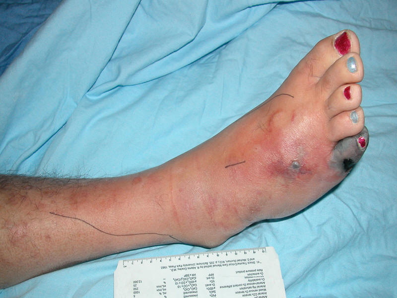
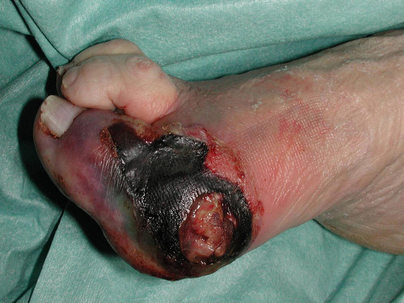
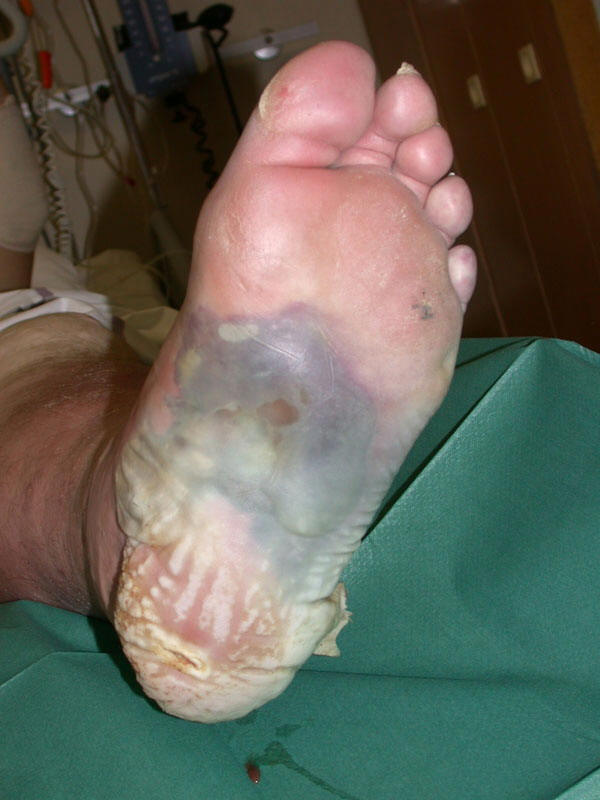
Moderate
![]() (III) to severe infections
(III) to severe infections
![]() (IV) often necessitate empirical regimens with activity
against commonly isolated gram-negative bacilli, MRSA (if there are risk factors
as previously discussed) and perhaps Enterococcus species. Enterococci
are relatively frequent isolates, especially in patients previously treated with
cephalosporins, but they are often colonizers and rarely primary pathogens.
Broadening empirical antibiotic coverage based on previously mentioned clinical
findings (Table 1) is often appropriate. As a general rule, we err on the side
of overly broad initial coverage, put our effort into obtaining high quality
specimens for culture, ensuring proper wound care and thinking more about the
definitive antibiotic regimen to complete the course when culture results are
available. We think most errors in treatment consist of failing to alter (and
curtail) antibiotic therapy rather than in making a poor initial antibiotic
choice.
(IV) often necessitate empirical regimens with activity
against commonly isolated gram-negative bacilli, MRSA (if there are risk factors
as previously discussed) and perhaps Enterococcus species. Enterococci
are relatively frequent isolates, especially in patients previously treated with
cephalosporins, but they are often colonizers and rarely primary pathogens.
Broadening empirical antibiotic coverage based on previously mentioned clinical
findings (Table 1) is often appropriate. As a general rule, we err on the side
of overly broad initial coverage, put our effort into obtaining high quality
specimens for culture, ensuring proper wound care and thinking more about the
definitive antibiotic regimen to complete the course when culture results are
available. We think most errors in treatment consist of failing to alter (and
curtail) antibiotic therapy rather than in making a poor initial antibiotic
choice.
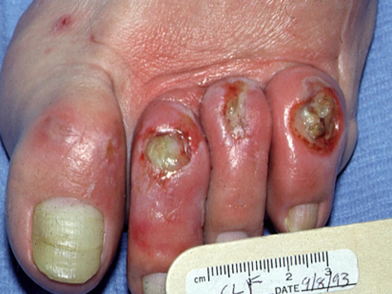
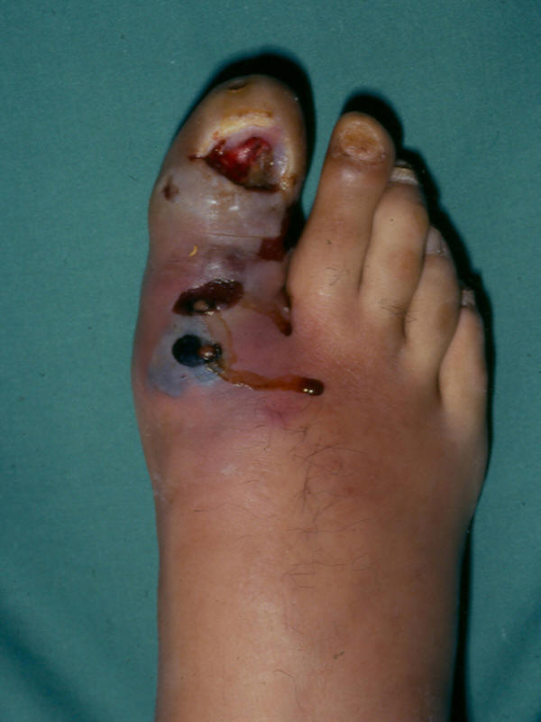
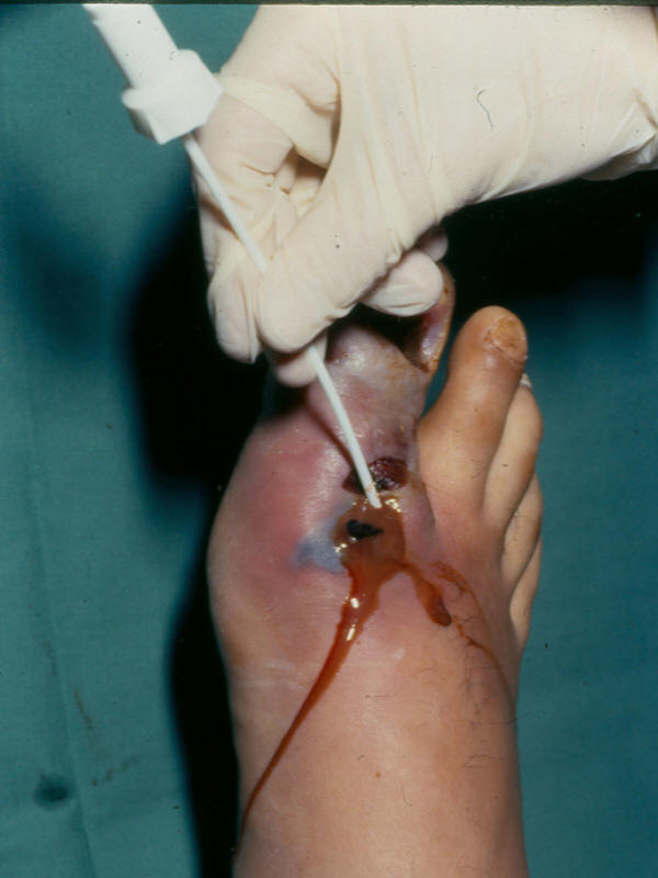
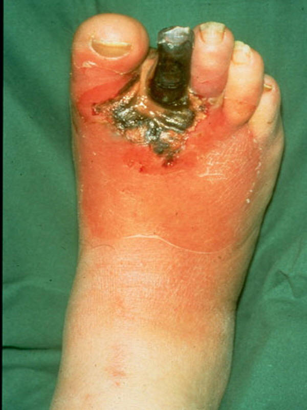
III.
| Moderate soft tissue infection of hammer toes | Septic great toe after surgery | Diabetic foot infection with gangrene | Necrotic toe and dorsal abscess from tight shoes | |
 |
 |
 |
 |
 |
IV.
| Moderate-to-severe diabetic foot infection with gangrene of the great toe | Severe diabetic foot infection | |
 |
 |
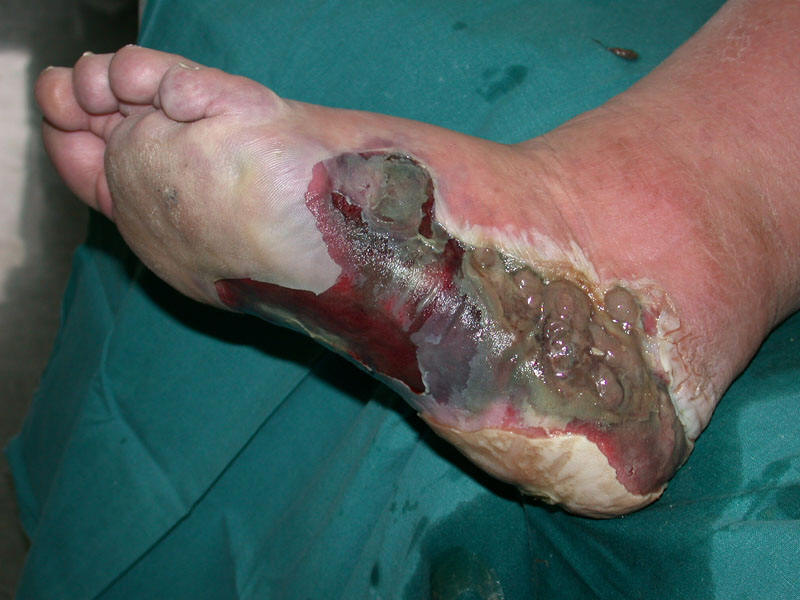 |
Table 1. Risk Factors for Foot Ulceration and Infection
|
Risk factor |
Mechanism leading to ulceration, impaired wound healing or infection |
|
Peripheral sensory neuropathy |
Loss of protective sensation (e.g., repetitive shear-type stress leading to ulceration) |
|
Peripheral motor neuropathy |
Abnormal foot anatomy and biomechanics resulting in excess pressure |
|
Peripheral autonomic neuropathy |
Impaired sweating leading to dry, cracked skin |
|
Arterial insufficiency |
Diminished delivery of nutrient, oxygen, neutrophils, etc. leading to impaired wound healing and clearance of infection |
|
Hyperglycemia |
Immune system (e.g., neutrophil) dysfunction and excess collagen cross-linking |
|
Patient disability or non-adherence |
Reduced vision (unable to inspect feet), prior amputation, lack of regular follow-up with medical care, poor hygiene, inappropriate footwear |
Additional considerations include the site of treatment and route of drug administration. Intravenous antibiotic therapy and hospitalization are not only costly but also potentially expose patients to iatrogenic complications and nosocomial infection. The severity of infection is usually the most important determinant of the need for hospitalization. Other factors may also influence the decision, such as the clinician’s desire to closely monitor the wound and response to treatment, a need for expedited diagnostic testing or consultant evaluation, and adverse social circumstances (e.g., inability of the patient to adequately care for the wound, off-load the foot, or uncertainty regarding compliance with antibiotics or follow-up care). Most patients with mild to moderate infections can be successfully treated as outpatients with oral antibiotic therapy. Many oral antibiotics have high bioavailability allowing therapeutic serum levels. An antibiotic’s pharmacokinetic and pharmacodynamic properties determine its serum, and therefore tissue, levels. When parenteral therapy is needed, it can often be offered by outpatient intravenous therapy with a once daily agent when this service is available. The arterial supply to the foot must also be adequate to ensure delivery of therapeutic antibiotic levels to the infected tissue.
After wound culture and antibiotic susceptibility results become available, the
clinician should consider narrowing the antibiotic coverage to the isolated
pathogen(s). Clinical assessment of the patient and wound, including the
response to the empirical regimen, are paramount in tailoring the antibiotic
regimen. If the wound is clinically responding, it is reasonable to continue an
empirical regimen, even if it does not have activity against all the isolated
organisms. Conversely, if a wound is not improving, broaden antibiotic coverage
to cover all isolated organisms, even those whose pathogenicity is uncertain.
The optimal duration of antibiotic therapy has not been studied, but we
recommend one to two weeks for mild to moderate soft tissue infections and at
least two weeks (occasionally longer) for deeper or more complex soft tissue
infections (Table 7). It is not usually necessary to continue antibiotic therapy
for soft tissue infections beyond the point when all clinical signs if infection
have resolved
![]() .
.
Debridement of an infected foot ulcer Probing of a diabetic foot ulcer after debridement
Table 7. Suggested Route, Setting, and Duration of Antibiotic Therapy by Clinical Syndrome
|
Site and severity of infection |
Route of antibiotic administration |
Usual setting for therapy |
Duration of therapy* |
|
Soft tissue only |
|||
|
Mild |
Oral |
Outpatient |
1-2 weeks, may extend up to 4 weeks if infection slow to resolve |
|
Moderate |
Oral (or initial parenteral) |
Outpatient or inpatient |
2-4 weeks |
|
Severe |
Initial parenteral, switch to oral when clinically stable |
Initial inpatient, discharge to outpatient when clinically stable |
2-4 weeks |
|
Bone or joint** |
|||
|
Post-amputation, no residual infected tissue |
Parenteral or oral |
Outpatient, unless patient ill or requires parenteral therapy (and no available OPAT) |
2-5 days |
|
Post-amputation, residual infected soft tissue but not infected bone |
Parenteral or oral |
|
2-4 weeks |
|
Post-amputation, residual infected but viable bone |
Initial parenteral, then consider switch to oral |
|
4-6 weeks |
|
Post-amputation, residual non-viable bone or no surgery |
Initial parenteral, then consider switch to oral |
|
>3 months |
OPAT = Outpatient antibiotic therapy
* Treat until resolution of all (or most) of the original symptoms and signs of infection
** With or without concurrent soft tissue infection
Surgical Intervention
Surgical procedures designed to control infection and preserve as much the
affected limb as possible are frequently required in the treatment of diabetic
foot infection. Early, thorough debridement of infected and necrotic tissue
helps define the extent of infection and may reduce the likelihood of future
amputation. Severe infections may be immediately limb-threatening and require
urgent surgical consultation. Unexplained, persistent foot pain, particularly in
a patient known to have advanced neuropathy (i.e., a previously insensate foot)
or distortion of the superficial anatomy (e.g., bulging of the plantar surface
![]() (V)
or dorsal induration
(V)
or dorsal induration
![]() (VI)
in a patient with a plantar ulcer) may indicate deep space
infection that warrants prompt evaluation by a surgeon. When amputation is
necessary, the surgeon should make every effort to perform a minor (below the
ankle) rather than a major amputation to preserve functionality. If severe
peripheral arterial disease is present, a vascular surgeon should be consulted
promptly. Non-urgent amputation should rarely be done without a vascular surgery
consultation. Early revascularization (i.e., within days) is generally preferred
to delaying the procedure for a prolonged course of potentially ineffective
antibiotic therapy. Endovascular or bypass procedures can be limb salvaging in
some patients with a diabetic foot infection.
(VI)
in a patient with a plantar ulcer) may indicate deep space
infection that warrants prompt evaluation by a surgeon. When amputation is
necessary, the surgeon should make every effort to perform a minor (below the
ankle) rather than a major amputation to preserve functionality. If severe
peripheral arterial disease is present, a vascular surgeon should be consulted
promptly. Non-urgent amputation should rarely be done without a vascular surgery
consultation. Early revascularization (i.e., within days) is generally preferred
to delaying the procedure for a prolonged course of potentially ineffective
antibiotic therapy. Endovascular or bypass procedures can be limb salvaging in
some patients with a diabetic foot infection.
V. Soft tissue abscess of 1st MTP area VI. Bulging foot abscess on the dorsal surface
Wound Care After the initial assessment and treatment, the patient requires ongoing wound care. Regular follow-up evaluations and repeated debridement of any residual dead tissue can help promote continued healing. Many dressings and wound-care products are marketed but none are favored based on evidence. A moist dressing that allows daily wound inspection is optimal. The roles of adjunctive treatments such as hyperbaric oxygen and granulocyte colony stimulating factors have not yet been adequately defined. Meta-analyses of these treatments suggest that an as yet undefined subset of patients may benefit from these treatments. Considering their cost they should rarely be used as initial therapy but rather reserved for patients who are not responding to what should otherwise be appropriate treatment.
Many clinicians and patients do not recognize the importance of off-loading pressure from the affected foot (1, 4). A variety of footwear devices are designed to redistribute pressure off the affected part of the foot. The total contact cast has been shown to be the best device, but the clinician and patient cannot easily view the infected wound in this device. In some patients healing sandals, half shoes, and removable cast walkers are adequate. Complete offloading of the affected limb may also be achieved with crutches, a walker or a wheelchair; however, these devices may increase the risk of ulceration of the contralateral limb and confinement to a wheelchair may not be practical and leads to deconditioning. With the exception of the total contact cast (which the patient cannot remove), patient compliance is often suboptimal. Select a dressing and off-loading device with the fact in mind that infected wounds must generally be examined and dressed daily.
Osteomyelitis
Osteomyelitis frequently complicates diabetic foot infection but its evaluation and management remain controversial. Except for direct inoculation by a puncture wound, bone infection spreads from soft tissue to cortex to marrow. Thus, nearly all cases in the setting of a diabetic foot infection are chronic osteomyelitis. Traditionally, surgical resection of infected and necrotic bone has been viewed as the best approach to curing these infections. Recent retrospective reports have demonstrated clinical success in ~65-80% of selected cases using prolonged (3-6 months) antibiotic (usually fluoroquinolone) therapy with little or no surgery. Prospective trials are needed to define in which clinical circumstances osteomyelitis may be appropriately treated without surgery. Using the results of bone culture is the preferred means to guide antibiotic therapy, especially if it is to be prolonged. When all the necrotic bone has been resected, 4-6 weeks of antibiotic therapy for the residual infected bone is usually sufficient. If nonsurgical treatment is selected, initial parenteral therapy (for perhaps 1-2 weeks) is recommended. Oral antibiotic(s) with good bioavailability may then be used to complete a course of therapy. If all infected and necrotic bone has been resected, a brief course of antibiotic therapy (1-2 weeks) for any residual soft tissue infection is sufficient. While much discussed, the data on the accuracy of measuring and importance of achieving adequate bone levels of antibiotics to treat osteomyelitis are weak. Some studies support the use of clindamycin; clinical experience suggests that beta-lactams and selected fluoroquinolones work well and some recent case series support using linezolid (for the shortest necessary duration and with careful monitoring for hematological and neurological toxicity).
PREVENTION
Having a diabetic foot infection increases the risk of future recurrence – emphasize this to the patient at this teachable moment to underscore the importance of preventive strategies. Identifying patients with peripheral neuropathy, foot deformity or peripheral arterial disease, and educating them about the risk of foot ulcer and diabetic foot infection is a simple, but critical and often overlooked, way to prevent diabetic foot infection. Primary and secondary prevention measures include optimizing metabolic (glycemic, lipid) control, wearing appropriate footwear, avoiding mechanical or thermal foot trauma, and performing a daily foot inspection. Fungal infections of the foot are more common in people with diabetes. Patients with tinea pedis should be treated with a topical anti-fungal, while those with severe onychomycosis may benefit from oral anti-fungal therapy. Persons with major foot complications should be referred to a specialist (e.g., podiatric, orthopedic or vascular surgeons).
CONCLUSIONS
Having a diabetic foot infection increases the risk of future recurrence – emphasize this to the patient at this teachable moment to underscore the importance of preventive strategies. Identifying patients with peripheral neuropathy, foot deformity or peripheral arterial disease, and educating them about the risk of foot ulcer and diabetic foot infection is a simple, but critical and often overlooked, way to prevent diabetic foot infection. Primary and secondary prevention measures include optimizing metabolic (glycemic, lipid) control, wearing appropriate footwear, avoiding mechanical or thermal foot trauma, and performing a daily foot inspection. Fungal infections of the foot are more common in people with diabetes. Patients with tinea pedis should be treated with a topical anti-fungal, while those with severe onychomycosis may benefit from oral anti-fungal therapy. Persons with major foot complications should be referred to a specialist (e.g., podiatric, orthopedic or vascular surgeons).