![]() Reprinted from
www.antimicrobe.org
Reprinted from
www.antimicrobe.org
Approach to the Patient with Fever and Rash
The differential diagnosis of acute fever and rash in an adult is quite extensive and includes a variety of infectious and non-infectious causes. Infectious disease emergencies and diseases that can have public health implications can present as fever and rash. Therefore, making a timely and specific diagnosis in an adult with fever and rash can be extremely important. An accurate characterization of the skin lesions and a thorough history can help narrow the differential diagnosis for a specific patient.
There are a limited number of skin manifestations that can result from a variety of insults including infection. The diagnosis can not be made by the description of the rash alone. There is no skin lesion that is pathognomonic for a specific pathogen. The skin manifestations associated with a specific infectious agent may be variable, and different types of skin lesions may be seen at different times in the course of one illness. Host factors such as neutropenia and thrombocytopenia may alter the appearance of the skin manifestations.
A variety of mechanisms may be involved in the pathogenesis of skin manifestations associated with infection. Skin lesions can develop secondary to multiplication of organisms within the skin. Organisms can produce toxins that can directly affect skin structure or can elicit an inflammatory response in the skin. Vascular involvement by the infection may also play a role. More than one mechanism may be involved in the development of a rash with a specific pathogen. Skin lesions involving the skin are referred to as exanthems and skin lesions involving mucous membranes are referred to as enanthems.
APPROACH TO THE PATIENT
Gather Data
A thorough history is extremely important in limiting the differential diagnosis for the adult with fever and rash (Table 1). Involvement in specific activities provides the potential for exposure to specific pathogens. Occupational exposures, contact with animals or ill individuals, ingestion of specific foods, sexual practices, and insect bites all may be important components of the history. A thorough medication history should be obtained including immunizations, over-the-counter preparations, past and present medications and any history of adverse effects related to any of these substances. Past medical history is important especially in regards to any immune compromise, valvular heart disease or splenectomy. Social history should include questions regarding intravenous drug use and travel. The time of year may be a vital consideration in the differential diagnosis.
Table 1. Important Historical Information in Evaluation of the Adult with Fever and Rash
Allergies/ Adverse effects Medications past and present
immunosuppressives
vaccines
over-the-counter medications Immune Status malignancy
transplantation splenectomy HIV Past medical history valvular heart disease renal failure diabetes mellitus liver disease trauma surgery Social History occupation travel (domestic and foreign) animal contact intravenous drug use ill contacts insect exposure sexual practices alcohol use dietary practices hobbies Rash where and when did it appear type of lesion pattern of spread associated symptoms timing of rash and associated symptoms pain or pruritus change in morphology of lesions Note – time of year and geographic location may also be important considerations
The history should also include specifics about the rash: where and when were the lesions first noticed, the nature of the lesions, the timing of the development of the skin findings with any associated symptoms, the pattern of spread or any change in the morphology of the lesions over the course of the illness, and if the lesions are painful or pruritic.
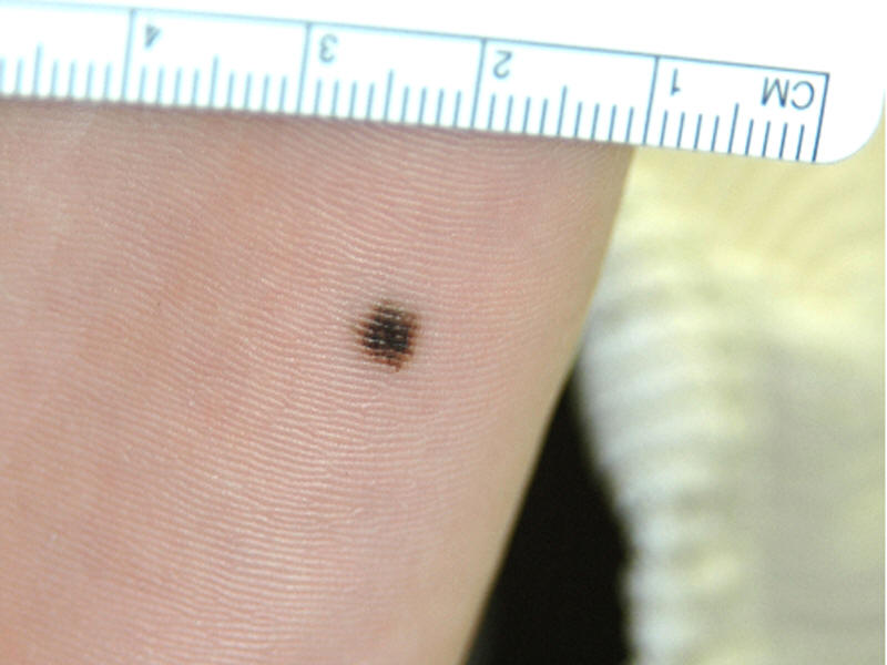
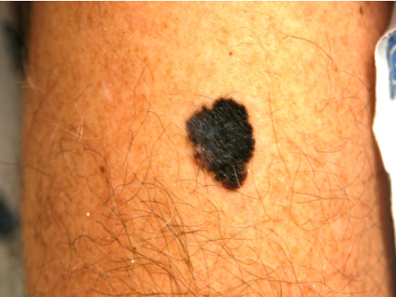
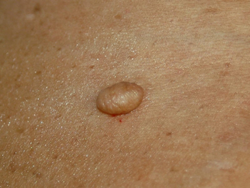
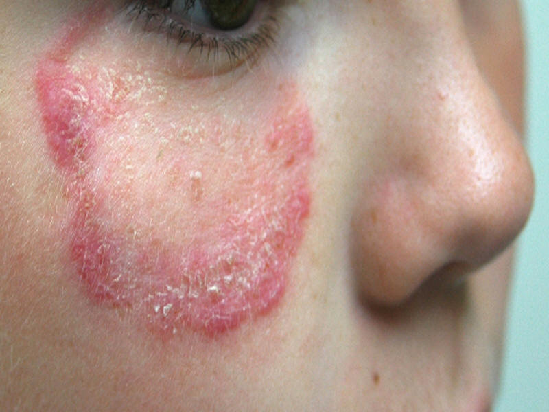
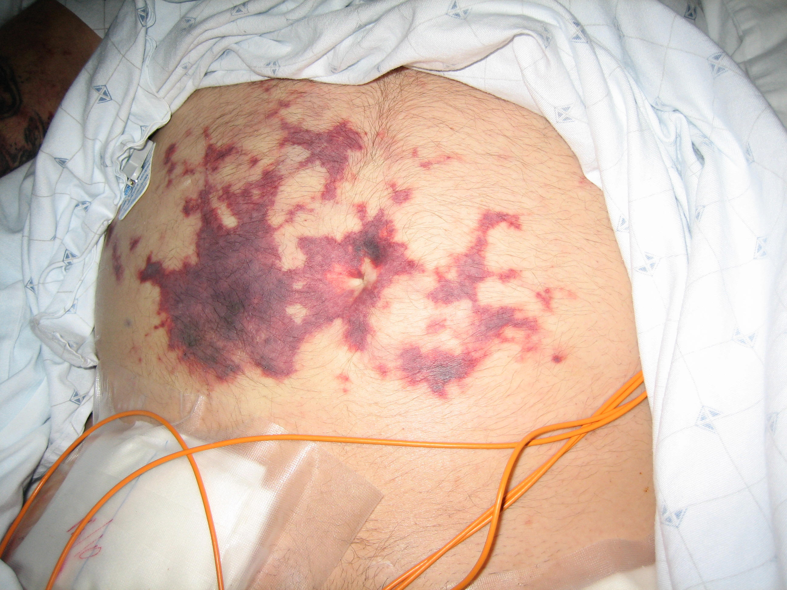
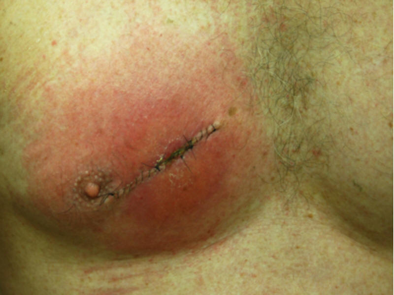
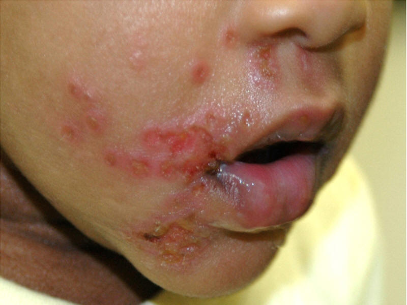
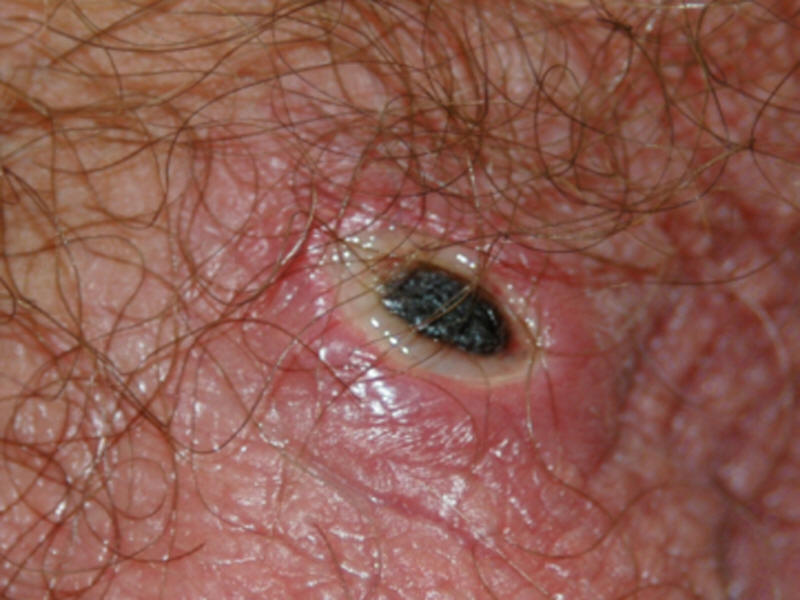
The differential for fever and rash in an adult is divided into categories based on the character of the predominant skin lesion. For the purposes of discussion in this chapter we will divide rash into six categories: macules/papules, diffuse erythema, vesicles/bullae, nodules, petechiae/purpura and urticaria. Therefore it is important that the skin findings are accurately characterized. Table 2 summarizes the common skin manifestations. A macule is a flat discoloration less than 1 centimeter in diameter. A common freckle would be an example of a macule. A patch is a large macule and a café au lait spot would be considered a patch. Macules and patches are generally not palpable. Papules are palpable, raised lesions less than 1 centimeter in diameter. Molluscum contagiosum is an example of an umbilicated papule. A plaque is a flat-topped raised lesion greater than 1.5 centimeters in diameter and the lesions of psoriasis can be described as plaques. Nodules are rounded, raised lesions greater than 1 centimeter in diameter. Tumors are usually greater than 2 to 3 centimeters in diameter. Vesicles are well circumscribed, fluid-filled lesions up to 1 centimeter in diameter and vesicles that are greater than 1 centimeter in diameter are classified as bullae. Pustules are elevated lesions filled with pus. A wheal or hive is a well-demarcated, elevated lesion that is usually pink in color and present for less than 24 hours. Petechiae and purpura represent bleeding into the skin. These lesions may or may not be palpable, but they do not blanche with pressure. Ecchymoses are large areas of bleeding into the skin. Certain infections can present with a diffuse erythema which blanches with minimal pressure. Crusts, scales, ulcers, and excoriations are secondary lesions that can complicate any of the primary skin lesions mentioned above.
Table 2. Types of Skin Lesions
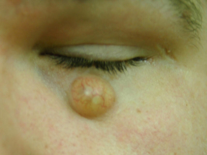
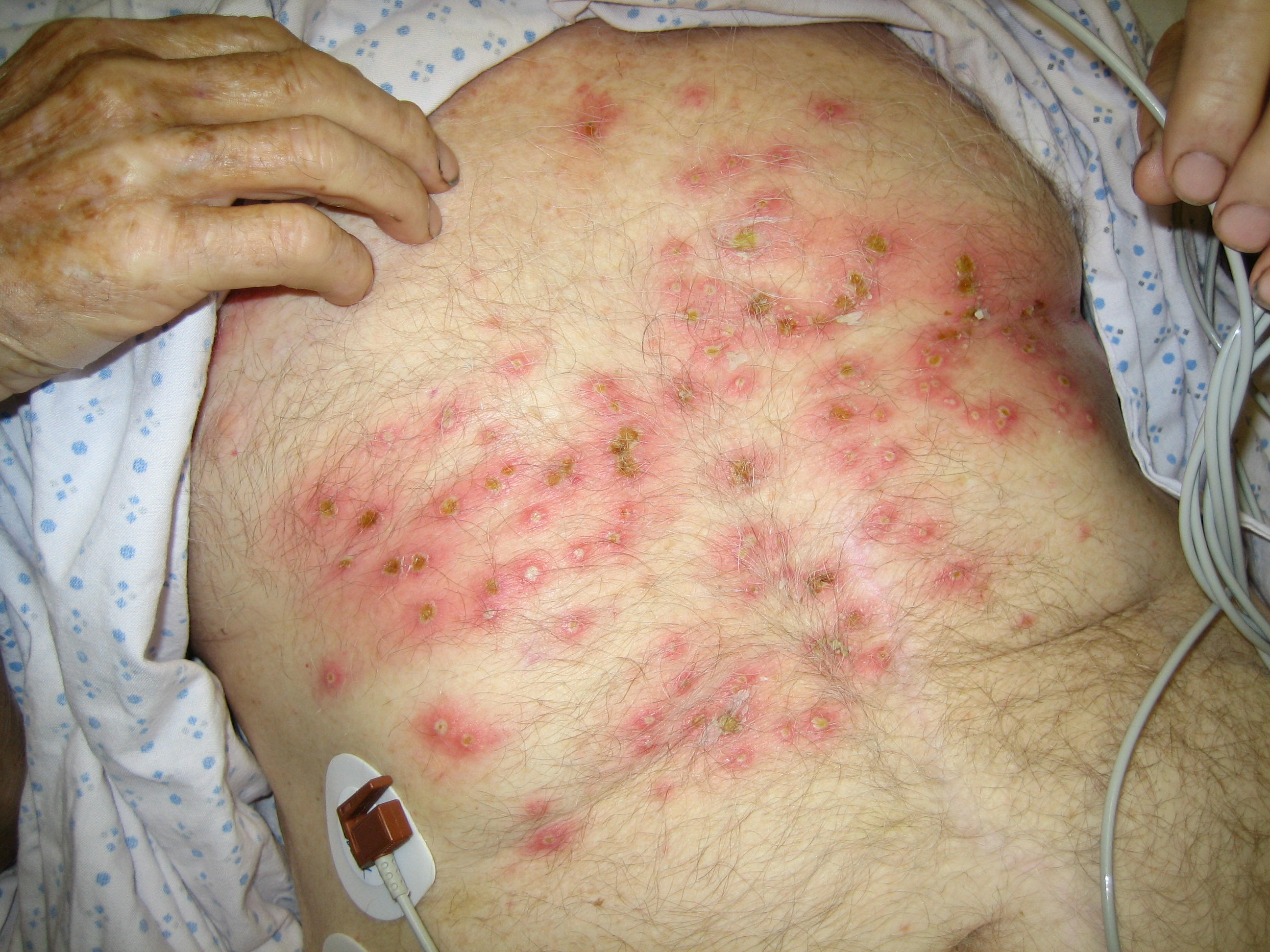
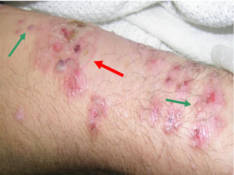
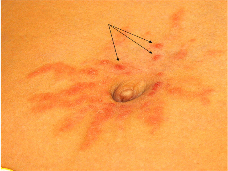
Macule – flat discoloration < 1 cm in diameter Patch – flat discoloration > 1 cm in diameter Papule – solid elevated lesion < 1 cm in diameter Plaque – flat-topped, elevated lesion > 1.5 cm in diameter Nodule – rounded, elevated lesion > 1 cm in diameter Vesicle – fluid-filled, elevated lesion up to 1 cm in diameter Vesicles (Green arrows)- Fluid-filled, elevated lesion less than 1 cm in diameter; Bulla (Red arrows)- Fluid filled, elevated lesions > 1 cm in diameter Pustule – pus-filled vesicle Wheal – well-demarcated, raised lesion lasting < 24 hours No Photo Petechia – pinpoint hemorrhage (does not blanche with pressure) Purpura – red macule/ papule that does not blanche with pressure Ecchymosis – large area of bleeding into the skin No Photo No Photo Erythema – redness that blanches with pressure Crust – dried exudate of blood and/ or plasma Eschar – hard crust or scab
When examining the patient with fever and rash it is important to examine the entire skin surface in good light, preferably natural light. The lesions should be palpated. As some skin lesions may be infectious it is advisable to wear gloves for the exam. In addition to accurate characterization of the skin lesions attention should be paid to the distribution and arrangement of the lesions. For example are the lesions grouped or linear in their arrangement. Mucous membranes in both the oral and genital regions, in addition to the hair, scalp, nails and intertriginous areas should all be examined. An assessment of the degree of illness of the patient is important. Patients who appear acutely ill or have unstable vitals signs may require urgent hospitalization and empiric therapy (Table 3). Other areas to emphasize on the physical examination are lymph nodes, liver and spleen, joints, and cardiac auscultation.
Table 3. Life-threatening Infections Presenting with Fever and Rash
Infection
Skin lesions
Historical clues
Diagnosis
Treatment
Isolation
Meningococcemia
petechiae
asplenia
Communal living
blood culture
CSF culture
Skin biopsy
Penicillin
ceftriaxone
yes
RMSF
petechiae
tick exposure
Spring
South central / midAtlantic US
serologyskin biopsy
doxycycline
no
Ehrlichia
maculopapular
tick exposure
Spring/ summer
serology
PCR
doxycycline
no
Capnocytophaga
petechiae
dog exposure
Asplenia
blood culture
penicillin
3rd gen ceph
no
Gram negative
Bacilli
papule, eschar
neutropenia
blood culture
anti-pseudomonal
beta lactam +/- AG
Carbapenem, Cipro
no
Vibrio
bullae
salt water exposure
Raw seafood
Chronic liver disease
blood culture
skin culture
surgery
doxycycline
cefotaxime, FQ
no
Toxic Shock Syndrome
erythema
tampon use
blood culture
skin culture
nafcillin
cefazolin, vanco
no
Necrotizing Fasciitis
erythema
bullae
trauma/ DM
obesity
tissue culture
blood culture
surgery
ABX based on site
no
Typhoid fever
maculopapular
international travel
blood culture
Stool culture
ceftriaxone
quinolone
no
Viral Hemorrhagic Fever
petechiae
international travel
serology
supportive
possible
Acute Endocarditis
petechiae
IVDU
valvular disease
blood culture
nafcillin or Vanco
no
RMSF = Rocky Mountain Spotted Fever, CSF = cerebral spinal fluid, PCR = polymerase chain reaction, AG = aminoglycoside, Cipro = ciprofloxacin, 3rd gen ceph = third generation cephalosporin, FQ = flouroquinolone, Vanco = vancomycin, DM = diabete mellitus, ABX = antibiotics
FEVER, RASH AND ARTHRITIS
Most of the syndromes that present with significant joint symptoms associated with fever and rash have already been mentioned in categorical sections (Table 11). Joints can be seeded during bacteremia associated with endocarditis or streptococcal toxic shock. Septic arthritis is more likely to occur in joints that are already damaged by prior trauma or chronic inflammation. Therefore it is important to sample the synovial fluid for culture in a patient with another potential cause for joint swelling and pain such as underlying Rheumatoid Arthritis.
Chronic meningococcemia presents as fever, rash and arthritis. The clinical picture is indistinguishable from that of disseminated Neisseria gonorrhoeae. The causative organism can be isolated from the blood.
Hepatitis B virus infection can present with rash (urticarial or maculopapular) and an oligo- or poly-arthritis. Transaminases may not be elevated at the time, but Hepatitis B sAg will be positive. Hepatitis C virus infection can also result in a rash and arthritis presentation which may or may not be associated with cryoglobulinemia.
Acute rheumatic fever (ARF) is a clinical syndrome that develops following a group A streptococcal (GAS) infection. Fever may be present but the major manifestations include migratory polyarthritis, carditis, subcutaneous nodules, chorea and the characteristic rash named erythema marginatum. The rash is erythematous but not painful or pruritic, and occurs predominately on the trunk and proximal extremities. The lesions are serpiginous with central clearing, and are fleeting on exam. The diagnosis of ARF is based on the clinical manifestations and evidence of recent GAS infection. Treatment is with anti-inflammatory medication.
Reiter's syndrome is a reactive arthritis that develops following certain gastrointestinal (Salmonella, Shigella, Campylobacter, Yersinia) or genitourinary (gonococcal or Chlamydia) infections. Although patients can have arthritis and rash, fever may not be present. Other associated findings include urethritis, oral ulcers, or ocular disease. A variety of collagen vascular diseases can have fever, rash, and arthritis as part of the clinical presentation.
Table 11. Fever, Rash and Arthritis
Infectious Chronic meningococcemia (Disseminated Neisseria gonorrhoeae ) Lyme (Borrelia burgdorferi) Rheumatic fever (GAS) German Measles (Rubella Virus) Parvovirus B 19 Endocarditis Hepatitis B or C Rate Bite fever (Streptobacillus moniliformis) HIV Non-infectious Juvenile Rheumatoid Arthritis Serum sickness Systemic Lupus Erythematosus Rheumatoid Arthritis
SUMMARY
This chapter has tried to provide a framework for the approach to a patient with fever and rash (Table 12). This chapter is not meant to be encyclopedic but has tried to highlight some of the common and important causes of fever and rash. Acute fever and rash in the adult can present a diagnostic challenge. One infectious agent can cause many dermatological syndromes and many agents can present with an identical dermatological picture. The key is to narrow the differential diagnosis by accurate characterization of the rash and a thorough history and physical examination. Once the differential is narrowed priority should be given to empiric therapy for patients who are unstable and may have a treatable, life-threatening infection. Isolation of certain patients with fever, or treatment of their contacts, may be important for public health reasons. Aggressive diagnostic modalities should be pursued especially in immunocompromised patients.
Table 12. Approach to Patient with Fever and Rash Obtain thorough history
Characterize rash
Assess for life-threatening illness
Consider differential diagnosis
Obtain diagnostic studies
Determine need for empiric antimicrobial therapy