Infectious conjunctivitis comes in two varieties: viral and bacterial (See Table 2). Viral conjunctivitis is more common and is typically adenoviral in etiology. Herpes simplex conjunctivitis is seen less frequently. The usual organisms implicated in bacterial conjunctivitis are Staphylococcus aureus, Staphylococcus epidermidis, Streptococcus pneumoniae, and Haemophilus influenza. An acute, hyperpurulent conjunctivitis is typically gonococcal. Chlamydia classically causes a chronic conjunctivitis.
Table 2. Viral vs. Bacterial Conjunctivitis
Viral Conjunctivitis
Bacterial Conjuncitivitis
Discharge
Watery, mucous
Purulent
Conjunctival pathology
Follicles
Papillae
Preauricular adenopathy
Common
Uncommon
Itching
Common
Less Common
Patients with conjunctivitis may have a clear history of recent contact with
individuals who had red eyes. The viral variety can be accompanied by an upper
respiratory infection or sore throat and typically begins in one eye and then
involves the second eye a few days later. A history of awakening with eyelids
stuck together is often elicited. General physical exam can reveal preauricular
lymphadenopathy, especially with the viral variety. Slit lamp examination can
help to distinguish between viral and bacterial causes. Conjunctival follicles
![]() are more commonly seen with viruses while papillae are seen in bacterial
conjunctivitis. There are exceptions to this rule, however, and often a mixed
picture with both papillae and follicles can be seen. Clear, mucoid discharge is
more indicative of a virus while purulent discharge is associated with bacteria.
For herpetic disease, finding periocular vesicles is a key to the diagnosis.
are more commonly seen with viruses while papillae are seen in bacterial
conjunctivitis. There are exceptions to this rule, however, and often a mixed
picture with both papillae and follicles can be seen. Clear, mucoid discharge is
more indicative of a virus while purulent discharge is associated with bacteria.
For herpetic disease, finding periocular vesicles is a key to the diagnosis.
|
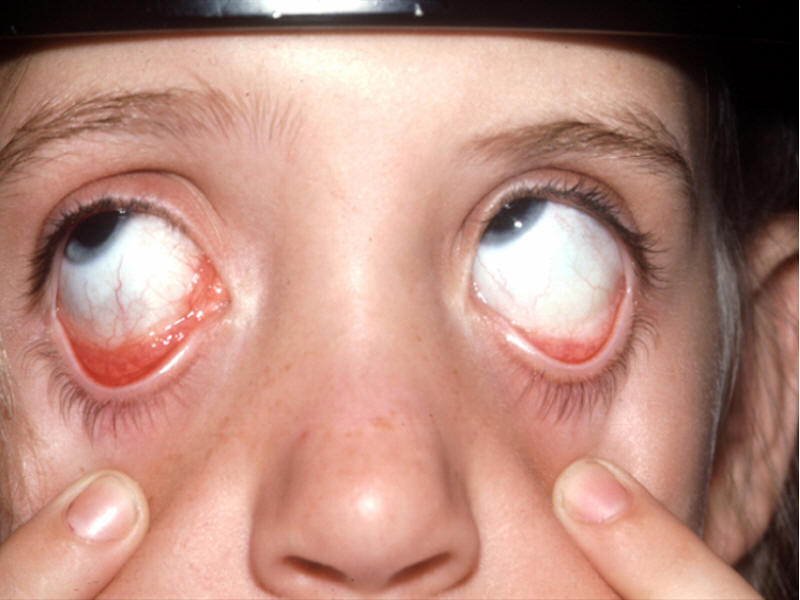
There is a follicular conjunctivitis of the right eye. The red “lumpy” appearance could be seen with adenoviral or allergic conjunctivitis. In this case it was due to infection with Bartonella henselae (cat-scratch disease). |
|
|
Clear cases of viral conjunctivitis do not require a work-up, but if there is any doubt as to the diagnosis, cultures of ocular discharge are required. Viral disease is treated conservatively with artificial tears and cold compresses. It often takes one to two weeks for symptoms to resolve. If the patient describes loss of vision, he or she should be referred to an ophthalmologist to ensure they have not developed subepithelial corneal infiltrates that require topical steroid therapy. Cases of herpetic conjunctivitis can be treated with vidarabine ointment four to six times per day.
Therapy of bacterial conjunctivitis should be guided by culture results. In general topical antibiotics are given for seven days. Some specific medications that cover most pathogens are moxifloxacin, gatifloxacin and polymyxin B/trimethoprim drops four to six times per day. Bacitracin and erythromycin ointment four times per day will also be sufficient in most cases. A few bacterial etiologies will require systemic medication. If the history or gram stain is suggestive of gonococcal disease, one gram of intramuscular ceftriaxone should be given promptly. If chlamydial disease is present, oral doxycycline or azithromycin therapy are acceptable therapies. Haemophilus influenzae conjunctivitis requires oral amoxicillin/clavunalate because of potential for concomitant nonocular involvement. With more virulent cases, especially gonococcal disease, referral to an ophthalmologist to rule out corneal involvement is imperative. Bacterial conjunctivitis should resolve within days of starting appropriate antibiotic therapy.

