Flow chart for
streptococci on Blood Agar Plate (BAP
)
BAP or SBA:
An abbreviation for blood agar
plate or sheep bloog agar. Blood agar contains mammalian blood (usually
sheep, rabbit or hrose), typically at a concentration of 5-10%. Blood
agar is an enriched media used to isolate bacteria and to detect
hemolytic activity.
PYR test: the PYR test is a qualitative
procedure for determining the ability of streptococci to enzymatically
hydrolyze L-pyrrolidonyl- β-napthylamide (PYR). A positive PYR tests
allows for the presumptive identification of group A streptococci (Streptococcus
pyogenes) and group D Enterococci.
Campy agar: Campylobacter CVA Agar is a selective
medium used in the primary isolation and cultivation of Campylobacter
jejuni from human fecal specimens. This selective medium contains
cefoperazone, vancomycin and amphotericin B; this combination of
antimicrobial agents inhibits the normal fecal flora for easier
detection of C. jejuni.
PYR test: the PYR test is a qualitative
procedure for determining the ability of streptococci to enzymatically
hydrolyze L-pyrrolidonyl- β-napthylamide (PYR). A positive PYR tests
allows for the presumptive identification of group A streptococci (Streptococcus
pyogenes) and group D Enterococci.
PYR test: the PYR test is a qualitative
procedure for determining the ability of streptococci to enzymatically
hydrolyze L-pyrrolidonyl- β-napthylamide (PYR). A positive PYR tests
allows for the presumptive identification of group A streptococci (Streptococcus
pyogenes) and group D Enterococci.
Bile Solubility: The bile solubility test is a
qualitative procedure for determining the ability of bacterial cells to
lyse in the presence of bile salts (sodium desoxycholate) under specific
conditions of time and temperature. The test is primarily used to
differentiate bile soluble Streptococcus pneumoniae from bile
insoluble alpha-hemolytic streptococci.
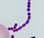
Gram stain: The Gram stain, is a
laboratory staining technique that distinguishes between two groups of
bacteria that have differences in the structure of their cell walls.
Standard bacterial taxonomy makes a distinction between Gram-negative
bacteria, which stain red/pink and the Gram-positive bacteria, which
stain blue/purple. Different antimicrobial agents are directed
specifically at gram-positive bacteria and gram-negative bacteria.
Catalase test: The catalase
test is used to differentiate some bacterial species. The test is done
by placing a drop of hydrogen peroxide on a microscope slide. Using an
applicator stick, a small portion of a colony is then added to a drop of
hydrogen peroxide drop.
If bubbles or froth forms, the organism is
said to be catalase-positive.
Staphylococci and micrococci are catalase-positive
If no bubbles form, the organism is catalase-negative.
Streptococci and Enterococci are catalase-negative
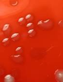
Beta hemolysis (β-hemolysis): Beta hemolysis is
the complete lysis of the red blood cells around and under the colonies
on a blood agar plate. This area appears transparent. Streptococcus
pyogenes displays beta hemolysis and is often called Group A
beta-hemolytic strep (GABHS).
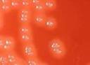
Alpha hemolysis (α-hemolysis):
Alpha hemolysis is the incomplete lysis of the red blood cells around
and under the colonies on a blood agar plate. This area appears dark and
greenish. Streptococcus pneumoniae and a group of streptococci (Streptococcus
viridans or viridans streptococci) found in oral flora display alpha
hemolysis.
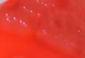
Non-hemolytic (γ-hemolysis): If an organism
does not induce any hemolysis on a blood agar plate, it is said to
display gamma or no hemolysis. The agar under and around the colony is
unchanged.