BAP or SBA: An abbreviation for blood agar plate or sheep blood agar. Blood agar contains mammalian blood (usually sheep, rabbit or horse), typically at a concentration of 5-10%. Blood agar is an enriched media used to isolate bacteria and to detect hemolytic activity.
Choc: Chocolate agar is a non-selective, enriched growth medium. It is a variant of the blood agar plate. It contains red blood cells, which have been lysed by heating. Chocolate agar is used for growing fastidious (hard to grow) bacteria.
N. mening grows on Choc & BAP
Catalase test: The catalase test is used to differentiate some bacterial species. The test is done by placing a drop of hydrogen peroxide on a microscope slide. Using an applicator stick, a small portion of a colony is then added to a drop of hydrogen peroxide drop.
  • If bubbles or froth forms, the organism is said to be catalase-positive.
    Staphylococci and micrococci are catalase-positive
  • If no bubbles form, the organism is catalase-negative. Streptococci and Enterococci are catalase-negative

  N. gonorrhoeae strong + catalase with 30% H2O2

  N. meningitidis weak few bubbles with 30% H2O2

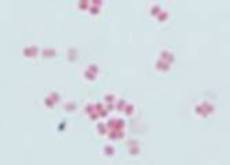
Neisseria meningitidis, N. gonorrhoeae, and Moraxella catarrhalis

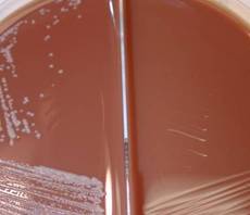
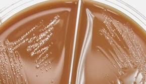
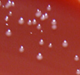
Chocolate, BAP, & Nutrient agar needed for good identification

@ Ellen Jo Baron 2007

  M. catarrhalis on  Choc NOT TM

Gram stain: The Gram stain, is a laboratory staining technique that distinguishes between two groups of bacteria that have differences in the structure of their cell walls. Standard bacterial taxonomy makes a distinction between Gram-negative bacteria, which stain red/pink and the Gram-positive bacteria, which stain blue/purple. Different antimicrobial agents are directed specifically at gram-positive bacteria and gram-negative bacteria.
Choc: Chocolate agar is a non-selective, enriched growth medium. It is a variant of the blood agar plate. It contains red blood cells, which have been lysed by heating. Chocolate agar is used for growing fastidious (hard to grow) bacteria.
BAP or SBA: An abbreviation for blood agar plate or sheep blood agar. Blood agar contains mammalian blood (usually sheep, rabbit or horse), typically at a concentration of 5-10%. Blood agar is an enriched media used to isolate bacteria and to detect hemolytic activity.

All 3 major gram neg diplococci grow well on chocolate agar
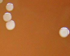
N. meningitidis and Moraxella catarrhalis grow on BAP and nutrient agar

M. catarrhalis grows at 22°C on BAP; N. meningitidis does not

  M. cat & N. men. grow on BAP
GC: GC is an abbreviation for gonococcus (Neisseria gonorrheoae).
TM: Tm is an abbreviation for Thayer Martin agar. Thayer Martin agar is used for the isolation of Neisseria species, including Neisseria gonorrhoeae and Neisseria meningitides; this medium inhibits the growth of most other microorganisms.
  GC & N. mening. on Choc & TM
GC: GC is an abbreviation for gonococcus (Neisseria gonorrheoae).
  GC or M. catarrhalis on Choc
Butyrate: Butyrate is a rapid test for the detection of the enzyme butyrate esterase in bacteria isolated on culture media. It is used for the presumptive identification of Moraxella (Branhamella) catarrhalis.
  M. catarrhalis is Butyrate +
Gram stain: The Gram stain, is a laboratory staining technique that distinguishes between two groups of bacteria that have differences in the structure of their cell walls. Standard bacterial taxonomy makes a distinction between Gram-negative bacteria, which stain red/pink and the Gram-positive bacteria, which stain blue/purple. Different antimicrobial agents are directed specifically at gram-positive bacteria and gram-negative bacteria.
Gram negative diplococci from colony
Oxidase: The oxidase test is used to determine if a bacterium produces certain cytochrome c oxidases. The reagent, tetramethyl-p-phenylenediamine is used as a redox indicator. The reagent turns dark blue when oxidized (oxidase positive). The reagent is colorless when reduced (oxidase-negative). Pseudomonas species and Aeromonas species are gram-negative bacilli that are oxidase-positive. Neisseria species are gram-negative cocci that are oxidase positive.
  All 3 species oxidase +
N. meningitidis in blood
Gram stain: The Gram stain, is a laboratory staining technique that distinguishes between two groups of bacteria that have differences in the structure of their cell walls. Standard bacterial taxonomy makes a distinction between Gram-negative bacteria, which stain red/pink and the Gram-positive bacteria, which stain blue/purple. Different antimicrobial agents are directed specifically at gram-positive bacteria and gram-negative bacteria.

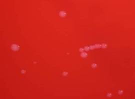
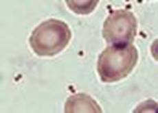
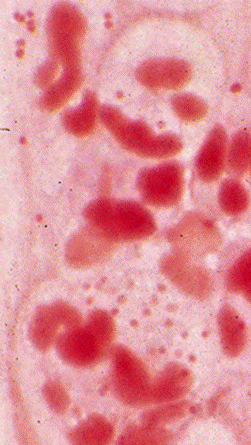
In original specimen:

Gram negative diplococci, some intracellular