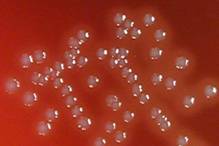
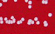
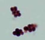
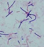
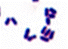
Cocci
Irregular, coryneform
Coagulase +
  Beta hemolytic
Beta hemolysis (β-hemolysis): Beta hemolysis is the complete lysis of the red blood cells around and under the colonies on a blood agar plate. This area appears transparent. Streptococcus pyogenes displays beta hemolysis and is often called Group A beta-hemolytic strep (GABHS).
Catalase -
Corynebacterium
Arcanobacterium

Listeria

Bacillus

Lactobacillus

Staphylococcus or

Micrococcus

Tube Coagulase test: The coagulase test is used to differentiate Staphylococcus aureus from coagulase-negative staphlycocci. The test uses rabbit plasma that has been inoculated with a staphylococcal colony. The tube is then incubated at 37 degrees Celsius for 1-1/2 hours. If negative, then incubation is continual up to 24 hours. This test detects free coagulase (coagulase that is released by bacterial cells into culture).
  • Positive (i.e., the suspect colony is S. aureus). The serum will coagulate, resulting in a clot (sometimes the clot is so pronounced that the liquid will completely solidify.
  • Negative, the plasma remains liquid. A negative result may be S. epidermidis.

  See Strep Chart

Flow chart Aerobic Gram positive organisms

@ Ellen Jo Baron 2007

Rods
Catalase test: The catalase test is used to differentiate some bacterial species. The test is done by placing a drop of hydrogen peroxide on a microscope slide. Using an applicator stick, a small portion of a colony is then added to a drop of hydrogen peroxide drop.
  • If bubbles or froth forms, the organism is said to be catalase-positive.
    Staphylococci and microcci are catalase-positive
  • If no bubbles form, the organism is catalase-negative. Streptococci and Enterococci are catalase-negative
 Beta hemolytic on Day 2
 Catalase negative
Beta hemolysis (β-hemolysis): Beta hemolysis is the complete lysis of the red blood cells around and under the colonies on a blood agar plate. This area appears transparent. Streptococcus pyogenes displays beta hemolysis and is often called Group A beta-hemolytic strep (GABHS).

Gram stain: The Gram stain, is a laboratory staining technique that distinguishes between two groups of bacteria that have differences in the structure of their cell walls. Standard bacterial taxonomy makes a distinction between Gram-negative bacteria, which stain red/pink and the Gram-positive bacteria, which stain blue/purple. Different antimicrobial agents are directed specifically at gram-positive bacteria and gram-negative bacteria.
Gram stain morphology
Regular rods
Yes
Yes
No
No
    Coagulase -
Coagulase: Coagulase is an enzyme produced by Staphylococcus aureus that converts fibrinogen to fibrin. In the laboratory, it is used to distinguish between different types of Staphylococci isolates. Coagulase negativity excludes S. aureus. S. aureus is coagulase-positive.


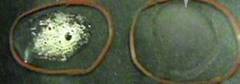
Slide coagulase test: The slide Coagulase test detects bound coagulase (clumping factor). This type of coagulase is attached ot the bacterial cell walls (surface). This test is usually performed on a glass slide.

Irregular, coryneform
      Catalase +
Catalase test: The catalase test is used to differentiate some bacterial species. The test is done by placing a drop of hydrogen peroxide on a microscope slide. Using an applicator stick, a small portion of a colony is then added to a drop of hydrogen peroxide drop.
  • If bubbles or froth forms, the organism is said to be catalase-positive.
    Staphylococci and microcci are catalase-positive
  • If no bubbles form, the organism is catalase-negative. Streptococci and Enterococci are catalase-negative